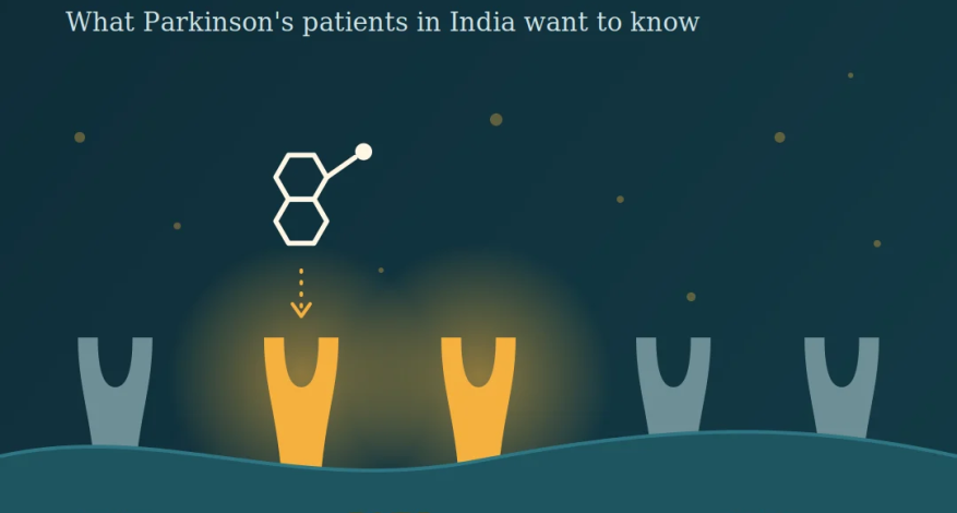
Illustration of the tavapadon molecule approaching D1/D5 dopamine receptors on a brain cell, beside the D2/D3 receptors targeted by older Parkinson’s drugs.news

Tavapadon (Juvmo): What Parkinson’s Patients and Families in India Want to Know

The 60-second version

  • What it is: Tavapadon, brand name Juvmo (AbbVie), is a once-daily tablet for Parkinson’s disease. It is the first approved medicine that switches on only the D1/D5 family of dopamine receptors.
  • What it does: It treats movement symptoms. It can be used alone in early Parkinson’s, or added to levodopa in people whose medicine wears off.
  • What it does not do: It has not been shown to slow or stop the disease. It is not a cure.
  • Where it stands: Approved by the US FDA in late September 2026, with US availability expected from October 2026.
  • India: [At the time of writing the product is not launched in India – October 2026] Please do not stop, change or add any Parkinson’s medicine on the strength of this page.
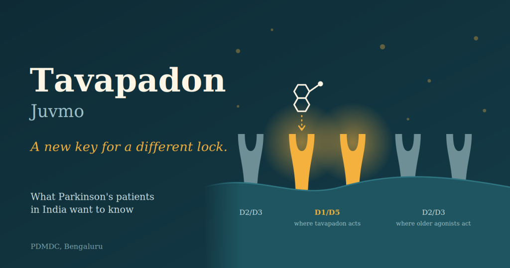
Illustration of the tavapadon molecule approaching D1/D5 dopamine receptors on a brain cell, beside the D2/D3 receptors targeted by older Parkinson’s drugs

Part 1: The basics

Why is everyone talking about tavapadon?

Because new types of Parkinson’s tablets are rare. Levodopa, the mainstay of treatment, dates back to the 1960s, and the dopamine agonists many patients take (pramipexole, ropinirole, rotigotine) all work on the same receptor family. Tavapadon takes a different route. The company describes it as the first dopamine-based breakthrough in decades, and neurologists have called it the first new class of dopamine agonist in a long time. That is enthusiasm worth taking seriously, but not at face value. See Part 3.

What is tavapadon, in plain language?

Parkinson’s happens because the brain cells that make dopamine, a chemical messenger that smooths movement, gradually die. Medicines help by topping up or imitating dopamine.

Tavapadon imitates dopamine. It was originally developed at Pfizer, later taken forward by Cerevel Therapeutics, and came to AbbVie when AbbVie acquired Cerevel in 2023.

What is special about its mechanism?

Dopamine docks onto several “locks” (receptors) in the brain. Think of two groups:

  • D2/D3 receptors: the target of today’s dopamine agonists. Switching these on helps movement, but is also linked to nausea, sleepiness, swelling, hallucinations and impulse-control problems.
  • D1/D5 receptors: the target of tavapadon. These also help movement, and the hope is that avoiding D2/D3 means fewer of those side effects.

Tavapadon is also a partial agonist. It switches the receptor on to a gentler degree than full dopamine would, which is intended to give steadier stimulation.

Honest caveat: this is the design rationale, not a proven advantage. No trial has yet compared tavapadon head-to-head with levodopa or with existing agonists.

How is it taken?

As a tablet by mouth, once a day, with or without levodopa. In the trials the dose was built up gradually over several weeks, with doses from 5 mg to 15 mg studied. Your own dose, if it is ever prescribed, would be set by your neurologist.


Part 2: What do the trials show?

Approval rests on three large phase 3 trials, called TEMPO-1, -2 and -3, plus a long open-label extension, TEMPO-4.

Trial Who Main result
TEMPO-1 (529 people) Early Parkinson’s, little or no prior treatment Combined movement and daily-living score improved by about 10 points on tavapadon versus a small worsening on placebo
TEMPO-2 (304 people) Early Parkinson’s, flexible dose About 9 points better than placebo
TEMPO-3 (507 people) On levodopa, with “wearing-off” About 1.9 hours less OFF time per day (versus 0.9 hours with placebo); “good ON” time rose by 1.7 hours (versus 0.6)

Scores are on the MDS-UPDRS Parts II and III, the standard clinical rating scale.

Is that a big improvement?

It is a real, statistically convincing benefit over placebo. In TEMPO-3, the extra benefit over placebo is roughly one additional hour of good ON time a day. For someone struggling with unpredictable OFF periods, an hour matters. It is, however, a modest gain and not a transformation.

What about the long term?

In the extension study, after about 85 weeks, roughly nine in ten participants had not needed to start levodopa (monotherapy group) or raise their levodopa dose (add-on group). These figures come from the company, have no comparison group, and only count people who stayed on the drug, so they should be read as encouraging rather than conclusive.


Part 3: Safety and side effects

What side effects were seen?

  • Used alone: nausea, headache, dizziness, tiredness, altered taste, vomiting, dry mouth, anxiety.
  • Used with levodopa: nausea, dyskinesia (involuntary movements), dizziness, headache, hallucinations, and a drop in blood pressure on standing.

Most side effects were mild to moderate. But in one early-Parkinson’s trial, about one in four people stopped tavapadon because of side effects (versus about one in twenty-five on placebo), mostly while the dose was being increased. Tolerability during titration is a genuine hurdle.

What warnings does the label carry?

The US prescribing information warns about low blood pressure on standing, hallucinations, dyskinesia, and impulse-control behaviours such as compulsive gambling or shopping. The last warning is shared with existing dopamine agonists. In other words, tavapadon does not escape every concern of its class.

Who has not been studied well?

People over 80 years were excluded from the pivotal trials. Evidence for very elderly patients, for people with significant other illnesses, and for real-world use is still thin.


Part 4: The Indian patient’s questions

Can I get it in India right now?

As of this writing [october 2026] the medications is not yet approved by India’s drug regulator (CDSCO) or launched here. Drugs are approved country by country, so a US approval does not mean Indian availability.

Should I buy it from overseas or online?

We advise against it. Medicines from unofficial online sellers may be counterfeit, wrongly dosed or stored badly. Importing an unapproved medicine can also be unlawful without permission. For a drug that needs careful dose build-up and monitoring, going without supervision carries real risk.

How much will it cost?

No Indian price has been announced that we could find. Levodopa-based therapy remains inexpensive in India, and several dopamine agonists, amantadine, MAO-B inhibitors and COMT inhibitors are already available. Any new branded drug is likely to be costlier at first, so cost will matter for many families in a long-term condition.

I am doing well on my current medicines. Should I switch?

No, not on your own, and not because a drug is new. If your current regimen controls your symptoms with tolerable side effects, “new” is not a reason to change. Never stop levodopa or an agonist suddenly, since abrupt withdrawal can be dangerous.

Who might be a candidate if it becomes available?

This is a question for your neurologist. The people the trials were designed for are:

  1. Adults in early Parkinson’s, as a first medicine.
  2. People on levodopa with wearing-off or fluctuations, as an add-on.

In practice, doctors will weigh your age, blood pressure, history of hallucinations or impulse-control problems, other medicines, and cost.

Does it slow the progression of Parkinson’s?

No evidence of that. Tavapadon relieves symptoms. Treatments aimed at slowing the disease remain under research.

Does it replace levodopa or deep brain stimulation (DBS)?

No. Levodopa remains the foundation of treatment, and tavapadon was tested alone or alongside it. DBS and other device-aided therapies are for a different stage and a different question.

Were Indian patients part of the evidence?

We have not seen India-specific results. Responses to medicines can differ between populations, so local experience will matter once the drug is in use here.


Part 5: Questions to bring to your neurologist

  1. Is my current treatment still working, or am I a candidate for something new?
  2. If tavapadon becomes available, would it replace a medicine, or be added to one?
  3. How would you build up the dose, and how will we handle nausea or dizziness along the way?
  4. Given my blood pressure and any past confusion or hallucinations, is it a safe fit for me?
  5. What will it cost per month, and for how long would I need it?
  6. What signs should make me call you urgently?

When to seek help promptly

Contact your doctor if you notice fainting or severe dizziness on standing, new hallucinations or confusion, new urges to gamble, shop or overspend, or sudden worsening of involuntary movements. This applies to any dopamine-acting Parkinson’s medicine.


The bottom line

Tavapadon is a genuinely new idea in Parkinson’s treatment: a once-daily tablet that targets a different dopamine receptor family and has solid placebo-controlled evidence for easing movement symptoms. It is not a cure, not proven better than what we already use, and not yet confirmed as available in India. For now, the best step is a conversation with your neurologist about whether your current treatment is serving you well.

Questions about your Parkinson’s care? Contact PDMDC on +91 70266 03300 or visit movementdisordersclinic.com.

This page is for general education and does not replace medical advice. Information reflects publicly reported FDA approval details and published trial results as of October 2026.

Sources: AbbVie press release (28 Sept 2026); TEMPO-1 (JAMA Neurology 2026), TEMPO-2 (Lancet Neurology 2026), TEMPO-3 (JAMA Neurology 2026); NeurologyLive and Medscape coverage of the FDA approval.

READ MORE
news

Parkinson’s Disease in 2025: 10 Scientific Breakthroughs That Are Changing the Future of Care

Parkinson’s disease is no longer viewed as a single, static condition—but as a complex, evolving brain disorder influenced by genetics, biology, lifestyle, and time. The year 2025 marked a turning point, with science moving beyond symptom control toward earlier diagnosis, smarter therapies, and the possibility of slowing disease progression. From stem-cell transplants and gene therapies to blood-based biomarkers and non-invasive brain treatments, research in 2025 expanded both the depth and breadth of how Parkinson’s is understood and managed.
This page highlights the Top 10 most important scientific developments in Parkinson’s disease in 2025, explained in a way that connects laboratory discoveries to real-world impact for patients, caregivers, and families—cutting through hype while focusing on what truly matters in care.

Top 10 Breakthroughs in Science for Parkinson's Disease in 2025

10) A next-generation once-daily dopamine agonist (tavapadon) was submitted for FDA review

Original sources: AbbVie NDA submission announcement and PD foundation coverage. AbbVie News Center+1
What it contained (scientific): Regulatory submission seeking approval for tavapadon—positioned as a once-daily oral option for PD (pending review outcomes). Michael J. Fox Foundation+1
Lay implication: The medication toolbox keeps expanding—sometimes the “win” is simpler dosing and smoother day control, not just brand-new mechanisms. Michael J. Fox Foundation+1
How it impacts PD care: If approved, it could offer another way to tailor therapy early and across progression—potentially improving adherence and symptom stability.

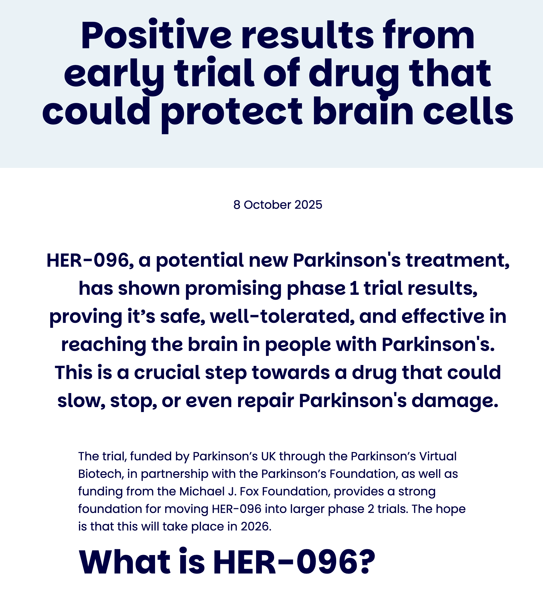
9) A new potential neuroprotective candidate (HER-096) reported encouraging early trial signals

Original source: Parkinson’s UK update (early trial results). Parkinson’s UK
What it contained (scientific): Phase 1 results emphasizing safety/tolerability and evidence the compound reaches the brain—foundational steps for any therapy aiming at neuroprotection/repair. Parkinson’s UK
Lay implication: Before we can prove a drug slows PD, we first prove it’s safe and actually gets to where it needs to act. Parkinson’s UK
How it impacts PD care: Not a clinic treatment yet, but it expands the pipeline of “protect the brain” approaches beyond dopamine replacement.

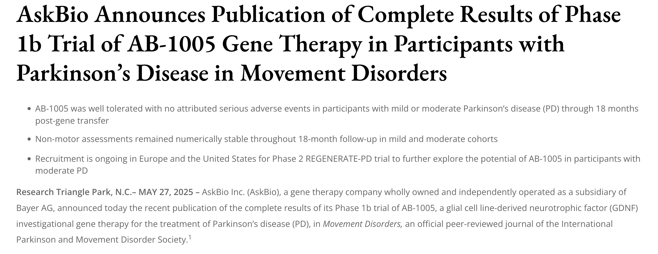
8) A gene-therapy program (AB-1005) published Phase 1b results and moved the field forward

Original source: AskBio publication announcement of Phase 1b AB-1005 gene therapy results in Movement Disorders (company summary). AskBio
What it contained (scientific): Early clinical data focusing on safety/tolerability and feasibility of gene therapy delivery; positioned as a potential approach to slow progression (still investigational). AskBio
Lay implication: Gene therapy for PD is no longer “science fiction”—but it’s still in the careful, step-by-step proof stage. AskBio
How it impacts PD care: Mostly through trial availability and accelerating know-how about dosing, delivery, and long-term monitoring.

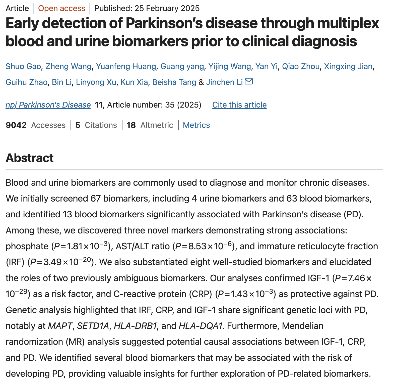
7) Another large biomarker push suggested panels (blood/urine markers) linked to PD risk years before diagnosis

Original science publication: Biomarker screening work in npj Parkinson’s Disease (2025). nature.com
What it contained (scientific): Screening many routine biomarkers, narrowing to a subset associated with PD, including several highlighted as strongly associated in that dataset. nature.com
Lay implication: PD risk may eventually be estimated using combinations of routine lab patterns—like “risk fingerprints,” not a single magic number. nature.com
How it impacts PD care: Supports the idea that future PD care may include risk staging (like cardiology risk scores), particularly for research enrollment and prevention trials.

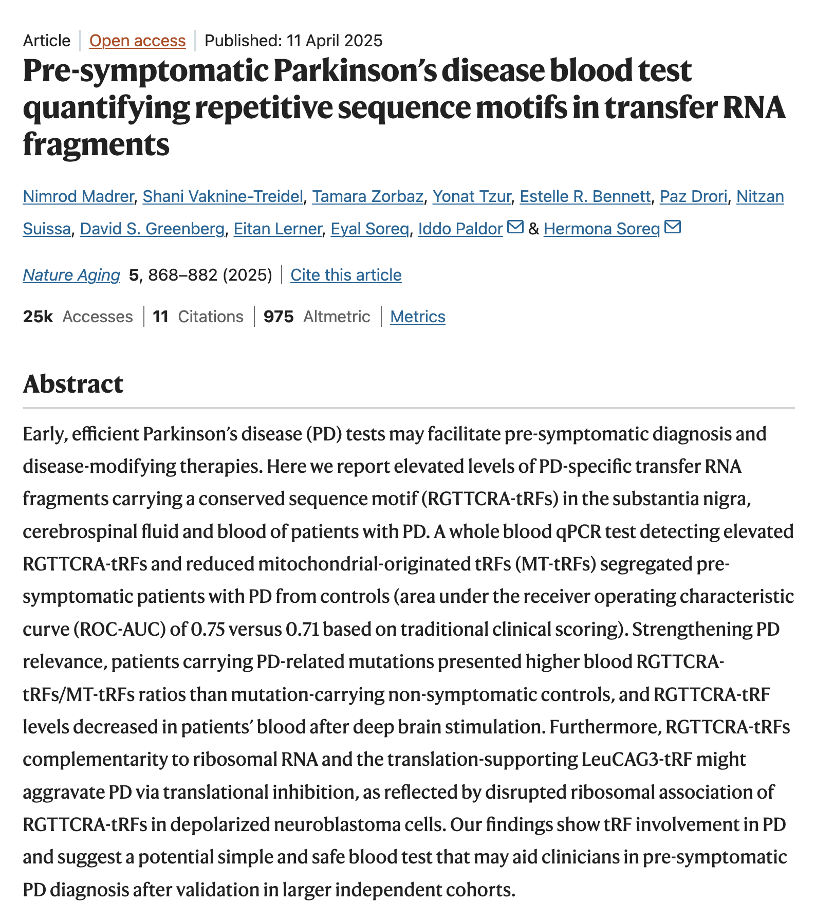
6) A blood-based RNA signal for very early PD detection was reported (pre-motor stage potential)

Original science publication / coverage: RNA/tRNA-fragment blood test work reported in Nature Aging (as covered by major PD organizations). American Parkinson Disease Association+1
What it contained (scientific): A method to detect PD-associated RNA fragment patterns in blood—aiming to identify PD before classic motor symptoms appear. American Parkinson Disease Association
Lay implication: This is part of the race toward a “blood test for Parkinson’s,” especially for early detection. American Parkinson Disease Association
How it impacts PD care: Not ready for routine clinic use, but it strengthens future pathways for:

  • screening high-risk people, and

  • getting patients into disease-modifying trials earlier.

5) Roche decided to take an anti-alpha-synuclein antibody (prasinezumab) forward into Phase 3

Original source: Roche announcement about advancing prasinezumab into Phase III based on trends and biomarker signals. Roche
What it contained (scientific): Reported signals (not a “cure claim”) suggesting reduced motor progression trends over 2 years in a study population and biomarker evidence consistent with target engagement/biology impact. Roche
Lay implication: A serious attempt at disease-modifying therapy (slowing progression), not just symptom relief, is being pushed into the definitive trial stage. Roche
How it impacts PD care: While not yet available clinically, it keeps the field focused on earlier diagnosis + earlier intervention strategies if/when such therapies prove effective.

4) A new continuous apomorphine infusion option got FDA approval for advanced PD “OFF” episodes

Original source: FDA approval announcement for ONAPGO (SPN-830) subcutaneous apomorphine infusion. Supernus+2Reuters+2
What it contained (scientific): Continuous subcutaneous delivery of apomorphine (dopamine agonist) aimed at reducing motor fluctuations; late-stage data showed meaningful reduction in daily OFF time vs placebo. Reuters+1
Lay implication: Instead of repeated rescue doses, medication can be delivered more steadily—helping reduce “wearing off.” Reuters
How it impacts PD care: More personalization for advanced PD—especially when oral absorption is erratic or OFF time dominates the day.

3) FDA cleared staged bilateral focused ultrasound for advanced PD symptoms (non-incision brain procedure)

Original announcement(s): FDA approval/clearance communications around staged bilateral focused ultrasound in advanced PD (foundation and manufacturer communications; clinical center explainer). Michael J. Fox Foundation+3FUS Foundation+3Insightec+3
What it contained (scientific): Expansion from one-sided to two-sided (staged) treatment using MRI-guided focused ultrasound to lesion a target pathway/region to reduce severe symptoms when medications fail. Insightec+1
Lay implication: A “no cut, no implant” option expanded—potentially helping people whose symptoms are disabling on both sides of the body. NewYork-Presbyterian+1
How it impacts PD care: Adds another advanced-therapy choice alongside DBS and infusion therapies—important for those who cannot (or do not want to) undergo implanted-device surgery.

2) A major stem-cell program moved into Phase 3 testing (closer to real-world approval pathways)

Original report: Bayer/BlueRock moved its PD cell therapy into Phase III trials (news report). Reuters
What it contained (scientific): Progression from earlier safety/tolerability and “cells functioning as intended” signals into the pivotal trial stage aimed at regulatory approval. Reuters
Lay implication: This is the step where treatments start being tested at the scale and rigor needed to become widely available. Reuters
How it impacts PD care: You may see more centers discussing trial eligibility, referral pathways, and long-term follow-up planning for restorative therapies.

1) Stem-cell dopamine neuron transplants showed encouraging early human results

Original science publication(s): Two clinical trials published in Nature (one using iPSC-derived dopamine progenitors; one using hES-derived dopamine neuron progenitors). nature.com+1
What it contained (scientific): Early-phase trials evaluating safety, cell survival, dopamine function (imaging evidence), and exploratory clinical changes after transplanting dopamine-producing precursor cells into the brain’s target regions. nature.com+2nature.com+2
Lay implication: “Replacing the missing dopamine cells” is moving from theory toward real-world feasibility—without tumors or major safety signals in these small studies. Parkinson’s Foundation+2nature.com+2
How it impacts PD care: Not a routine treatment yet, but it meaningfully strengthens the case that cell replacement could become a future option for selected patients—especially when symptoms are no longer well controlled by tablets alone.

To Conclude

Taken together, the breakthroughs of 2025 tell an important story: Parkinson’s care is moving from a reactive, symptom-based approach toward a future that is predictive, personalized, and potentially disease-modifying. While many of these advances are still in clinical trials and not yet part of routine treatment, they collectively reshape how Parkinson’s is diagnosed, monitored, and treated across its full journey. For patients and families, this means more informed choices, earlier intervention opportunities, and a growing pipeline of therapies aimed not just at living with Parkinson’s—but living better with it. Science does not change care overnight, but 2025 has clearly laid stronger foundations for the years ahead.

Acknowledgment
The identification of scientific publications and developments summarized on this page was assisted by AI-based search and aggregation tools to scan the 2025 Parkinson’s disease literature landscape. All shortlisted studies were manually reviewed, interpreted, and contextually evaluated by the author, and the final selection of the “Top 10” breakthroughs was made solely by the author based on scientific rigor, clinical relevance, and potential impact on patient care.


READ MORE
Awareness

History of Wilson’s Disease: How S.A.K. Wilson’s Discovery Transformed Movement Disorders

History of Wilson’s Disease: How S.A.K. Wilson’s Discovery Transformed Movement Disorders

From a fatal childhood mystery to a treatable genetic disorder – and the birth of movement disorders neurology.
By Dr. Prashanth LK · Parkinson’s Disease & Movement Disorders Clinic, Bangalore
Reading time: ~10 minutes

Samuel Alexander Kinnier Wilson (born 6 December 1878), whose work on “progressive lenticular degeneration” reshaped neurology.
At the start of the 20th century, neurology understood paralysis, stroke, and epilepsy. But patients with tremor, twisting postures, drooling, and emotional outbursts were medically homeless. Their illnesses had no clear name, no anatomic map, and certainly no effective treatment.That changed with a young neurologist born on 6 December 1878 – Samuel Alexander Kinnier Wilson. His work would not only define a new disease, now known as Wilson’s disease, but also give birth to the concept of the extrapyramidal system and eventually to a whole subspecialty: Movement Disorders.

“With a single, monumental paper in 1912, Kinnier Wilson showed the world that tremor, rigidity, and dystonia came from hidden motor circuits deep within the brain.”

Before Wilson: When Tremor Had No Map

In the early 1900s, doctors could localise weakness to the pyramidal tract, explain seizures as cortical storms, and recognise classical strokes. But they struggled to understand patients who:

  • Shook violently without weakness,
  • Developed bizarre grimacing and drooling,
  • Lost their speech, yet understood everything,
  • Showed behaviour changes and emotional lability,
  • And died young, often within the same family.

These patients were variously labelled as hysterical, degenerative, or simply “incurable.” The idea that deep brain circuits could generate complex abnormal movements – without paralysis – did not yet exist.

The 1912 Breakthrough: Progressive Lenticular Degeneration

In 1912, working at the National Hospital, Queen Square in London, Kinnier Wilson published a 214-page tour de force in the journal Brain titled:

“Progressive lenticular degeneration: a familial nervous disease associated with cirrhosis of the liver.”

He meticulously described four young patients, from overlapping families, who developed:

  • Generalised tremor and rigidity,
  • Severe dysarthria, drooling, and swallowing difficulty,
  • Abnormal postures and contractures,
  • Emotionalism and subtle mental changes,
  • And, at autopsy, silent but advanced cirrhosis of the liver.

At post-mortem, he found striking bilateral degeneration of the lenticular nucleus, part of what we now recognise as the basal ganglia. For the first time, a disease was shown to selectively attack deep motor circuits, producing profound movement abnormalities without primary pyramidal weakness.

“Progressive lenticular degeneration… is essentially and chiefly a disease of the extrapyramidal motor system.” – S.A.K. Wilson, 1912

Birth of the Extrapyramidal System

Wilson’s observations forced neurology to redraw its motor map. Until then, movement was largely equated with the corticospinal (pyramidal) system. Everything else – tremor, rigidity, dystonia, chorea – sat in an uncomfortable grey zone.

By carefully correlating clinical signs with lenticular pathology, Wilson argued that there must be:

  • A parallel motor network outside the pyramidal tract,
  • Responsible for tone, posture, and automatic movements,
  • Capable of producing abnormal movements without paralysis.

Thus emerged the concept of the extrapyramidal system. Over subsequent decades, this idea became the organising framework for conditions such as Parkinson’s disease, Huntington’s disease, dystonia, and tics – and laid the intellectual foundation for today’s movement disorders neurology.

The Cruel Reality: A Brilliant Description, But No Cure

Although Wilson’s clinico-pathological work was revolutionary, he confronted a heartbreaking truth: there was no treatment. Every patient with progressive lenticular degeneration deteriorated and ultimately died, often in adolescence or young adulthood.

Wilson himself longed for an effective therapy but passed away in 1937, decades before the disease that now bears his name became treatable.

From Autopsy Table to Copper: The First Clues

In 1913, just a year after Wilson’s paper, a pathologist reported increased copper in the liver of a patient with the same clinical picture. The observation was remarkable – but largely ignored.

For the next 30 years, scattered reports linked copper deposition to this mysterious disorder, yet the dots were not fully connected. The disease remained uniformly fatal.

The Copper Revolution: John Nathaniel Cumings

A major turning point came in 1948, when John Nathaniel Cumings demonstrated that patients with Wilson’s disease had massive copper accumulation in both the liver and brain. This firmly established the illness as a disorder of copper metabolism.Cumings proposed a daring idea: if copper was the toxin, perhaps it could be removed. He turned to a wartime antidote – British Anti-Lewisite (BAL), a chelating agent developed during World War II as a treatment for arsenical gas exposure.

Early trials showed that BAL could indeed mobilise copper and provide some neurological improvement. For the first time, Wilson’s disease was no longer absolutely fatal – but treatment required painful injections, carried significant toxicity, and had diminishing returns with repeated courses.

Penicillamine: A Serendipitous Eureka Moment

The true therapeutic breakthrough arrived in the 1950s through the work of John Michael Walshe. While studying amino acid metabolism in liver disease, Walshe identified a curious sulfur-containing compound in the urine of patients receiving penicillin – D-penicillamine.

Later, while working in Boston, he saw a young man with Wilson’s disease who was deteriorating despite BAL therapy. Walking back from the ward, Walshe had a sudden insight: penicillamine’s structure suggested that it might chelate copper even more efficiently.

In the ethics-light 1950s, he followed a simple rule: never give a patient a drug you are not willing to take yourself. He first took a gram of penicillamine, experienced no ill effect, and then administered it to the patient.

Copper excretion in the urine surged. Over time, the patient’s tremor softened and function improved. Subsequent series confirmed that oral penicillamine could transform Wilson’s disease from a lethal illness into a controllable, chronic condition.

“Once 100% fatal, Wilson’s disease is now one of neurology’s most treatable genetic disorders – provided it is recognised early and decoppering therapy is maintained.”

The Molecular Key: Ceruloplasmin and ATP7B

Parallel advances in biochemistry identified a copper-carrying plasma protein, ceruloplasmin, which was found to be low or absent in patients with Wilson’s disease. Later, molecular genetics pinpointed the causative gene, ATP7B, a copper-transporting ATPase.

Together, these discoveries completed the journey from clinical syndrome to molecular diagnosis: Wilson’s disease is now recognised as an autosomal recessive disorder of copper transport, affecting both liver and brain, but eminently treatable if detected early.

Wilson’s Disease at a Glance

  • Autosomal recessive genetic disorder of copper metabolism.
  • Involves both liver and brain; may also affect kidneys and bones.
  • Kayser–Fleischer rings and low ceruloplasmin are important clues.
  • Treatment: decoppering agents (e.g., penicillamine, trientine) and zinc.
  • With early, lifelong therapy, many patients can live near-normal lives.

India as a Hotspot

  • Significant clinical series from Indian centres have highlighted a high burden of Wilson’s disease.
  • Patients often present young, with severe neurological manifestations.
  • Misdiagnosis as psychiatric, epileptic, or other neurodegenerative disorders is common.
  • Awareness and early screening of siblings can dramatically change outcomes.

From One Rare Disease to a Whole Subspecialty

Wilson’s original description did much more than define a single rare disease. By showing that movement disorders could arise from specific basal ganglia pathology, he helped reshape thinking about Parkinson’s disease, Huntington’s disease, dystonias, and many other conditions.Over the 20th century, this conceptual framework evolved into a full-fledged subspecialty: Movement Disorders Neurology. Treatments such as levodopa therapy, botulinum toxin for dystonia, and deep brain stimulation for Parkinson’s disease all rest on the foundation that Wilson’s meticulous clinico-pathological work helped to build.

“Kinnier Wilson was the father of basal ganglia research. We stand on his shoulders.” – Marsden & Fahn

December 6: More Than a Birthday

Today, Wilson’s disease stands as one of medicine’s most remarkable transformation stories. Once uniformly fatal, it is now a condition in which – with early diagnosis and lifelong decoppering therapy – many patients can complete education, work, marry, and lead productive lives.

Each year, 6 December, the birthday of S.A.K. Wilson, offers an opportunity to remember:

  • The power of careful clinical observation,
  • The value of linking anatomy to physiology and biochemistry,
  • And the hope that rare, devastating diseases can become treatable.

In clinics around the world – including here in India – teenagers once bound to wheelchairs now walk, children once mute now speak, and families once resigned to tragedy now see a future. All trace, in some way, back to a young neurologist who refused to accept unexplained movement as a mystery.

If You Suspect Wilson’s Disease: What Next?

Warning Signs That Should Raise Suspicion
  • Unexplained tremor, stiffness, or abnormal postures in a child or young adult.
  • Behavioural change or psychiatric symptoms with liver problems or jaundice.
  • Family history of “mysterious” neurological illness or early-onset liver failure.

Wilson’s disease is treatable. Early diagnosis can prevent irreversible brain damage and chronic disability. If these features are present – especially in younger individuals – a focused evaluation for Wilson’s disease should be considered.

Patients and families in India who are concerned about Wilson’s disease can seek specialised assessment at dedicated Movement Disorders clinics, including the Parkinson’s Disease & Movement Disorders Clinic in Bangalore.

References & Further Reading

  • S.A.K. Wilson. Progressive lenticular degeneration: a familial nervous disease associated with cirrhosis of the liver. Brain. 1912.
  • Walshe JM. History of Wilson disease: a personal account. In: Handbook of Clinical Neurology – Wilson Disease. 2017.

  • Walshe JM. The conquest of Wilson’s disease. Brain. 2009.
  • Cumings JN. The copper and iron content of brain and liver in the normal and in hepatolenticular degeneration. Brain. 1948.
  • Lees AJ. John Michael Walshe – Obituary. Movement Disorders. 2023.
  • Prashanth LK et al. Wilson’s disease: diagnostic errors and clinical implications. J Neurol Neurosurg Psychiatry. 2004.

Last updated: December 2025


READ MORE
Chorea

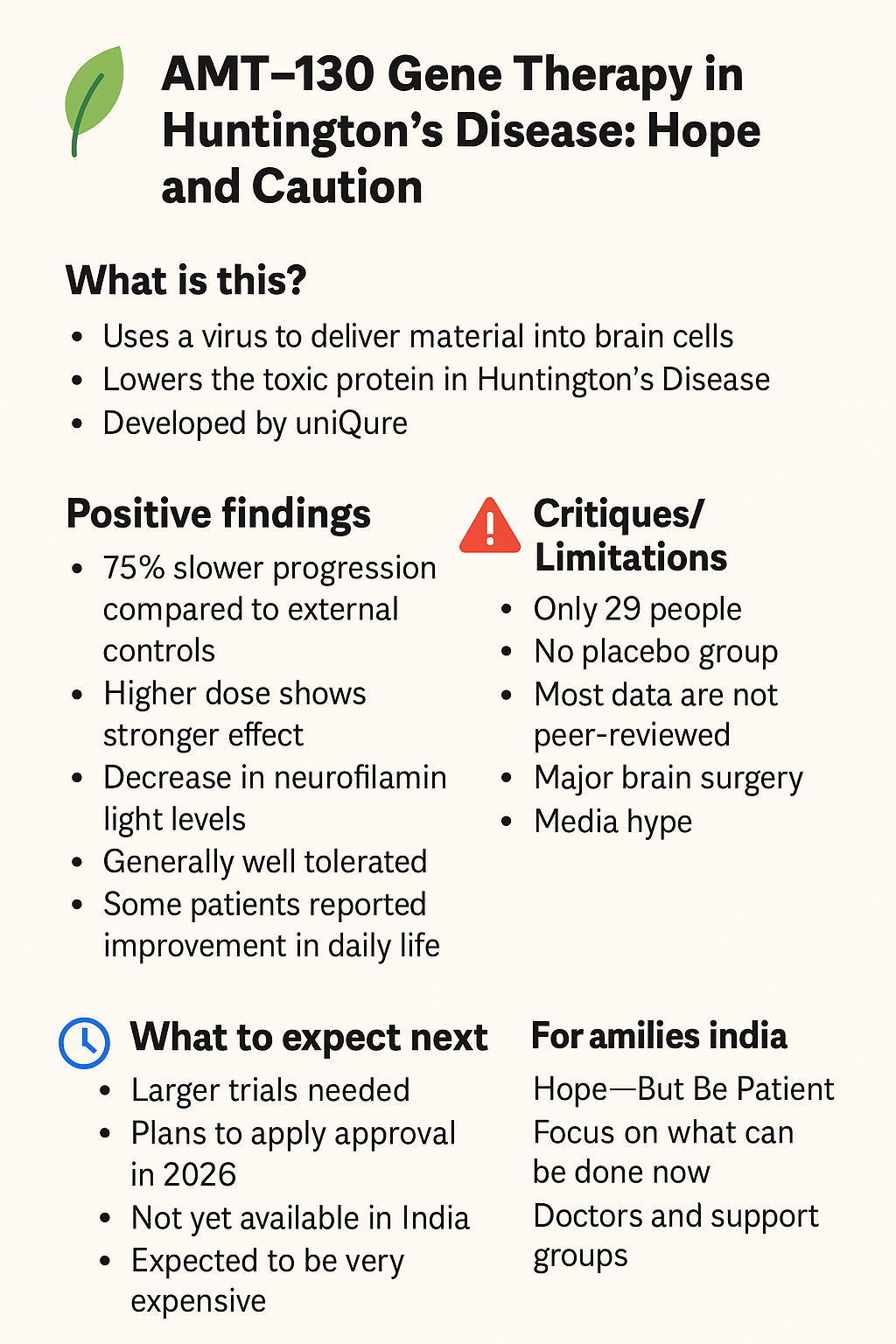
Huntington’s Disease in India: AMT-130 Gene Therapy Explained – Hope, Caution, and Costs

Introduction
Huntington’s disease (HD) is a rare, inherited brain disorder that affects thousands of families worldwide, including in India. It causes problems with movement, memory, and daily function, and currently has no cure.

In late 2025, news outlets and scientific groups around the world reported that a new experimental treatment — AMT-130 gene therapy — showed signs of slowing down the disease by up to 75% in a small clinical trial. Headlines have raised huge hopes. But what do these numbers mean? What are the positives, what are the limitations, and when could this treatment reach India?

This page explains AMT-130 in a Question & Answer format, using simple language for families.

Questions and Answers

1. What is AMT-130 gene therapy?

  • AMT-130 is the first gene therapy tested in humans with Huntington’s disease.

  • It was developed by uniQure, a biotech company based in the Netherlands and USA.

  • The goal: reduce the toxic huntingtin protein that damages brain cells in HD.


2. How does it work?

  • A harmless virus (called AAV5 vector) carries a small genetic code into brain cells.

  • This code produces micro-RNA, which acts like a “mute button” for the faulty huntingtin gene.

  • It stops brain cells from making as much toxic protein.

  • The therapy is given once, through 12–18 hours of neurosurgery, directly into brain areas called the caudate and putamen.

3. How was the study done?

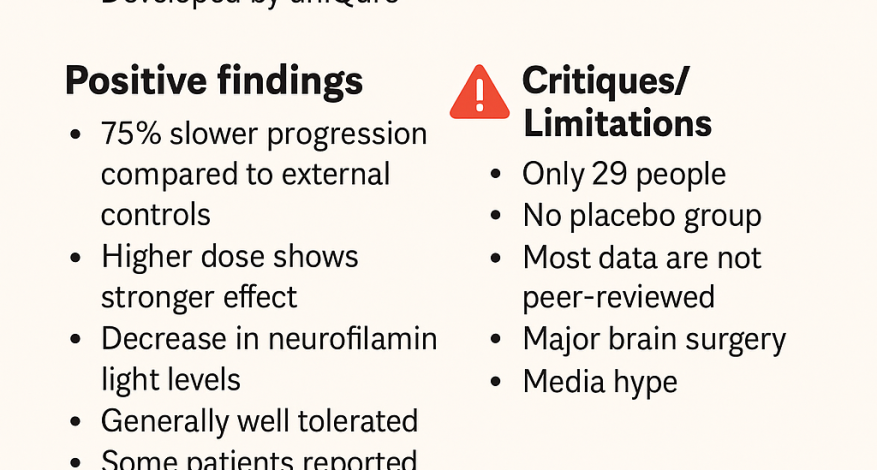
  • 29 patients with early Huntington’s disease joined the trial (low-dose and high-dose groups).

  • No placebo group was used. Instead, results were compared with historical/external controls (past patients with similar stage and age).

  • Patients are being followed for up to 36 months (3 years).


4. What are the positive findings?

✅ Slower decline:

  • At 36 months, decline on the cUHDRS scale was –0.38 points in treated patients vs –1.52 in controls → reported as 75% slower progression.

  • On another scale (TFC), decline was slowed by ~60%.

✅ Cognitive benefits:

  • ~88% slower decline in processing speed (SDMT test).

  • ~113% slower decline in Stroop word reading.

✅ Biomarkers:

  • CSF NfL (a marker of nerve injury) decreased by ~8.2% instead of increasing.

  • Early data suggested mutant huntingtin protein levels dropped by ~53.8% in some patients.

✅ Dose effect:

  • The higher dose group did better than the low dose group.

✅ Daily life impact:

  • Some patients reported better function — one returned to work, others were still walking when a wheelchair was expected.

✅ Safety:

  • Side effects (like inflammation and headaches) were manageable. No new major risks after 2022.


5. What are the critiques and limitations?

⚠️ Small trial: Only 29 patients, with just 12 in the high-dose group followed fully.
⚠️ No placebo group: Comparisons were against past data, not new untreated patients.
⚠️ Short-term issues: At 12 months, placebo/external controls looked better on some measures.
⚠️ Small effect size: On cUHDRS, the change (0.4 points in 3 years) may not be felt in daily life.
⚠️ Not peer-reviewed yet: Results mostly shared via company press releases and news.
⚠️ Surgery required: Long brain surgery with risks, so not a simple treatment.
⚠️ Cost and access: Gene therapies can cost over USD $2 million (~₹16–17 crore) per patient.


6. What does “75% slower progression” mean in real life?

  • It does not mean the disease is stopped or cured.

  • It means that, on paper, decline was slower than in comparison patients.

  • For example: what usually happens in 1 year might take 4 years.

  • This is encouraging, but based on small numbers and early data.

  • Some of the experts have been critical of this 75% highlight and have discussed this on their social media handles. Quoting one of the experts interpretation on Twitter / X platform : “Primary outcome measure: the composite Unified Huntington’s Disease Rating Scale (cUHDRS). This is a measure of overall disability (0–25; lower = worse). Typical range: ~2–18. Results at 36 months: AMT-130 group: −0.38 Historical cohort: −1.52 The company called this a “75% slower progression.” Here’s how they got it: (−0.38 −(−1.52))/−1.52 × 100 = 75% · However, this 75% figure represents a relative difference compared to a propensity-matched historical cohort — not a within-patient change, nor a clinically meaningful effect on the cUHDRS scale itself. In short: it sounds big, but it isn’t) Here are the key caveats 👇 1️⃣ Small numbers: Only 12 participants completed treatment in the high-dose group (out of 17). Data for the low-dose group (n=12) weren’t reported. 2️⃣ Small effect size: The average decline in cUHDRS in early HD is ~0.9–1.0 points/year. A 0.4-point change over 3 years (0.13/year) is below the level patients can perceive as meaningful. 3️⃣ No concurrent placebo: The “75%” comparison is vs. a historical cohort, not a placebo group. Given the invasive protocol (brain infusion + repeated lumbar punctures), placebo effects could be substantial. 4️⃣ High variability, weak comparison: The cUHDRS has large standard deviations driven by practice and placebo effects. That variability can inflate small open-label trials when compared to natural history data.“

7. What happens next?

  • Regulatory steps: uniQure plans to apply to the US FDA in 2026. Europe and UK will follow.

  • Peer-reviewed publications are expected soon.

  • Larger trials with proper placebo groups are needed.

  • India: Availability will take several years. Access will depend on approvals, pricing, and neurosurgical facilities.


💰 Cost and Access Issues

  • Similar gene therapies (for haemophilia, spinal muscular atrophy) cost ₹16–20 crore per patient.

  • AMT-130 may be in this range or higher.

  • Because it requires specialised brain surgery, access will be limited to major centres even in high-income countries.

  • In India, widespread access will be a major challenge without strong public health or philanthropic support.


🌸 Final Words for Indian Families

AMT-130 is a milestone — the first time a gene therapy has shown signals of slowing Huntington’s disease. It brings hope but must be understood with caution:

  • Early trial, small numbers.

  • Headlines can exaggerate.

  • Costs will be very high.

For now, families in India should:

  • Stay updated through support groups and doctors.

  • Participate in registries and observational studies.

  • Hold on to hope — but avoid false promises.

Interesting Related Videos / Informations

READ MORE
Awareness

#KeepMovingForParkinson: A Progressive Exercise Guide for People with Parkinson’s Disease

Welcome to the official movement guide from the #KeepMovingForParkinson initiative – a curated set of 16 functional exercises, categorized across 4 progressive levels of physical activity. This program is not time-bound. Instead, it is designed for people with Parkinson’s Disease to choose exercises based on their current mobility level – from bed-bound to fully mobile.

Each level contains 4 exercises, with clear steps, benefits, and guidance on who should perform them. Whether you’re starting from bed or standing strong, these movements are designed to help you stay active, build confidence, and improve quality of life.


🛎️ Level 1: Bed-Based Exercises

For individuals with limited mobility

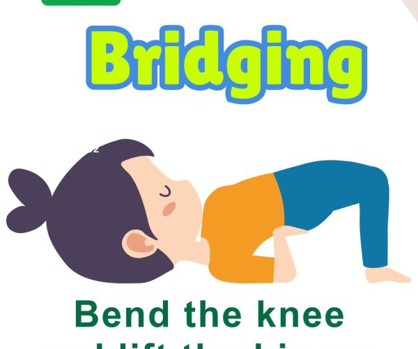
1. Bridging

Steps: Lie on your back with knees bent and feet flat. Slowly lift your hips upward. Hold for 10 seconds, lower back down. Benefits: Improves glute and core strength, reduces stiffness. Who Should Do It: Patients in bed rest or significant mobility limitation.


2. Butterfly Stretch

Steps: Sit or lie down, bring the soles of your feet together, knees out. Gently press knees toward the bed. Benefits: Enhances hip flexibility. Who Should Do It: Those with hip stiffness or reduced pelvic mobility.

3. Side-to-Side Leg Roll

Steps: Lie on your back with knees bent. Gently roll knees side to side. Benefits: Improves trunk rotation and spinal mobility. Who Should Do It: People with trunk stiffness or poor rotational movement.


4. Leg Raise

Steps: Keep one leg bent, other straight. Lift straight leg to 30 degrees. Hold, then lower. Benefits: Strengthens quads and core. Who Should Do It: Anyone needing lower limb activation from bed.

💺 Level 2: Chair-Based Exercises

For individuals able to sit upright with or without support.

5. Trunk Rotation

Steps: Sit tall, arms crossed over chest. Gently rotate the trunk side to side. Benefits: Improves spinal mobility and trunk control. Who Should Do It: Those beginning seated rehab.

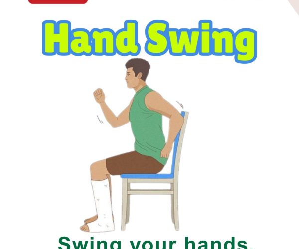
6. Hand Swing

Steps: Sit comfortably. Swing one arm forward and back rhythmically, then switch. Benefits: Promotes upper limb rhythm and arm swing. Who Should Do It: People with freezing or reduced arm movement.

7. Seated Marching

Steps: Lift one knee up at a time in a marching motion while seated. Benefits: Boosts leg coordination and circulation. Who Should Do It: Anyone preparing for standing transitions.

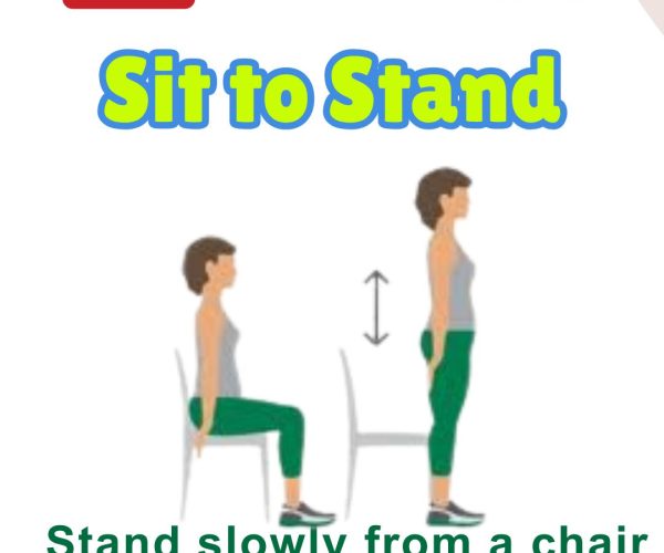
8. Sit to Stand

Steps: From seated position, rise to stand using as little hand support as possible. Benefits: Builds leg strength and functional independence. Who Should Do It: Ideal for improving transition from sitting to standing.

🧶 Level 3: Sit/Stand-Based Exercises

For individuals able to stand with support or partial independence.

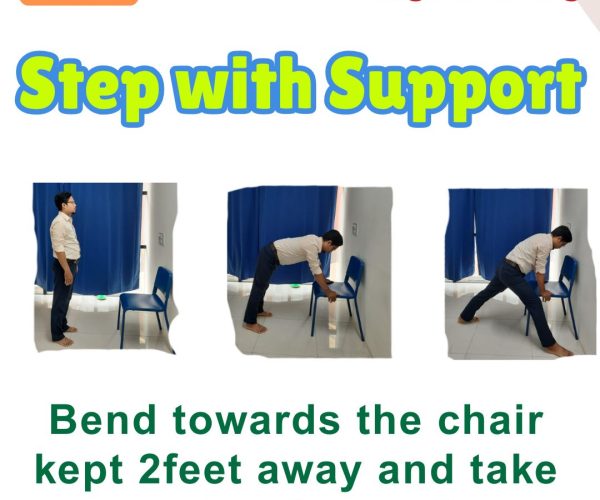
12. Step with Support

Steps: Hold a chair for support. Step forward and back slowly. Benefits: Builds stepping confidence. Who Should Do It: Anyone practicing gait with balance limitations.

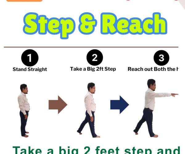
11. Step and Reach

Steps: Take a 2-foot forward step, reaching arms forward. Benefits: Improves stride length and coordination. Who Should Do It: For those retraining functional walking.

10. Side-to-Side Reach

Steps: Stand or sit. Reach one arm to the opposite side, alternating slowly. Benefits: Enhances trunk flexibility and balance. Who Should Do It: Patients working on side stability.

9. Floor to Ceiling Reach

Steps: Start seated or standing. Reach arms down to the floor, then up to the ceiling. Benefits: Encourages full-body range of motion. Who Should Do It: Those with mild movement initiation chall

🏃️ Level 4: Standing and Dynamic Balance Exercises

For individuals with good standing balance or progressing toward full mobility.

15. Rock & Reach

Steps: Stand tall, swing arms sideways and rotate the body. Benefits: Activates large movements, improves balance. Who Should Do It: Great for those practicing functional trunk rotation.

14. Hurdle Crossing

Steps: Step over low objects spaced 1 foot apart (use support if needed). Benefits: Trains high stepping and obstacle avoidance. Who Should Do It: People with reduced foot clearance.

13. Big Step Walk

Steps: Take long, deliberate steps around a room (goal: 2000 steps daily). Benefits: Improves gait and step length. Who Should Do It: Those with shuffling or freezing gait.

16. One Leg Stance

Steps: Stand and lift one leg off the ground. Hold for 10–15 seconds. Alternate. Benefits: Enhances single-leg balance. Who Should Do It: Suitable for fall-prevention and high-level balance training.

Final Notes

Consult your doctor or physiotherapist before starting new exercises.

Modify based on fatigue or stiffness.

Repeat exercises daily or on alternate days based on tolerance.

Patient Videos

READ MORE
Awareness

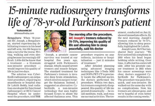
CyberKnife Radiosurgery Explained: Clarifying the 15-Minute Parkinson’s Procedure in the News

A recent news article highlighted a 78-year-old Parkinson’s disease patient whose life reportedly changed after a 15-minute CyberKnife radiosurgery session. Are you wondering how this procedure works, how it compares to other lesioning surgeries, or who might benefit from it?

In this video, we clarify the details behind the headlines (Cyberknife Radiosurgery for Parkinson’s) :

  • What is CyberKnife radiosurgery and what are Lesioning surgeries for the Brain?

    • Understanding this noninvasive, high-precision radiation treatment.
    • Also understanding various types of Brain Lesioning surgeries – Advantages and Limitations
  • How can it help Parkinson’s patients?

    • The mechanism behind targeting specific brain areas to reduce tremor and other movement symptoms.
  • Comparing different lesioning approaches

    • Focused Ultrasound (FUS), Gamma Knife, Radiofrequency ablation, and Deep Brain Stimulation (DBS)—what’s the difference?
  • Important considerations

    • Pros and cons, potential side effects, time to see results, and long-term follow-up.

We created this presentation to answer common questions from patients, caregivers, and anyone curious about newer treatments for Parkinson’s disease. This is not a replacement for medical advice; we encourage you to consult a movement disorder specialist or neurosurgeon to see if radiosurgery is right for you.

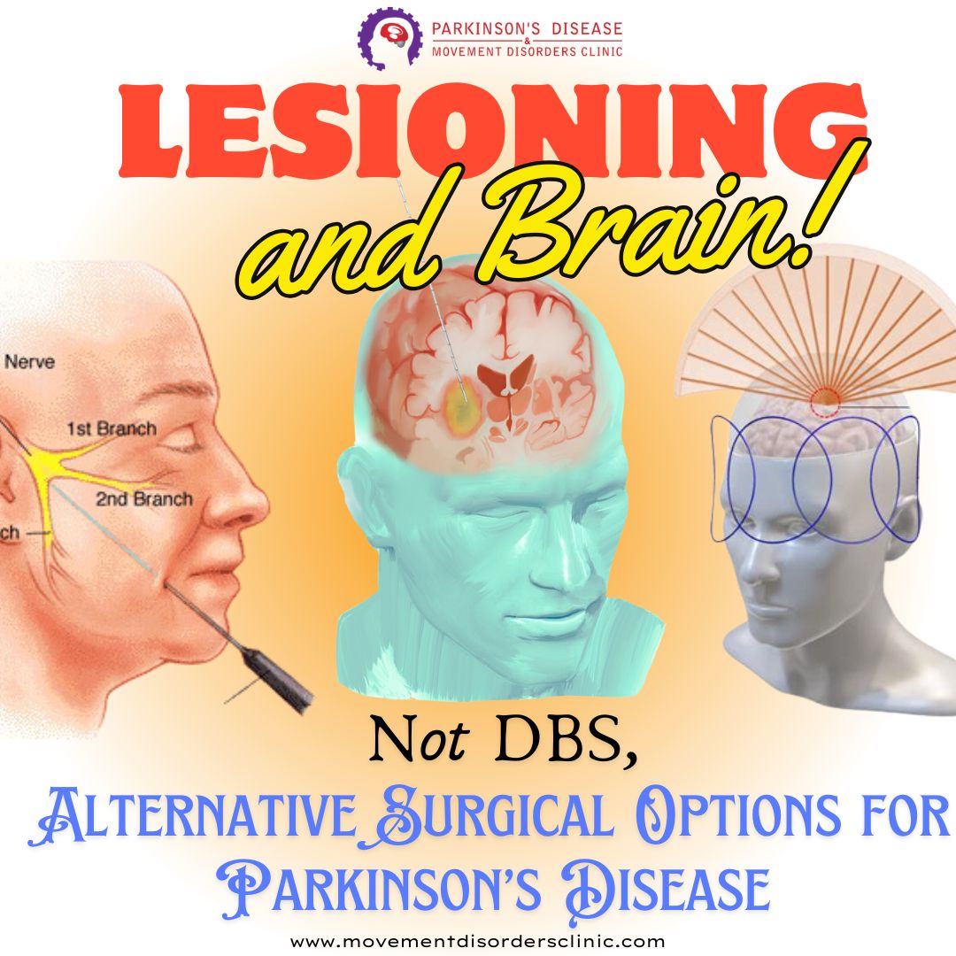
1. What Are Lesioning Surgeries?

Lesioning surgeries involve the use of techniques (chemical, radiofrequency, focused ultrasound, or even stereotactic radiosurgery) to create a precisely targeted lesion in a specific brain area thought to be responsible for a particular set of symptoms. By destroying these targeted cells or interrupting pathological circuit activity, symptom relief can often be achieved.

Common Techniques for Creating Lesions

  • Radiofrequency Lesioning: A probe is inserted stereotactically, and high-frequency currents produce heat that destroys the target tissue.
  • Focused Ultrasound (FUS): Uses high-intensity ultrasound waves focused on a specific area in the brain without needing an incision, guided by MRI.
  • Stereotactic Radiosurgery (e.g., Gamma Knife): Uses focused radiation beams to lesion the target gradually.
  • Chemical Lesioning (much less common today): Injecting chemicals that destroy local neurons.

2. Historical Context

  1. Early Psychosurgery (e.g., Lobotomies)

    • In the early to mid-20th century, surgeries like the prefrontal lobotomy were used to treat psychiatric disorders, often with severe and unpredictable side effects. Because of ethical concerns and poor patient outcomes, these procedures largely fell out of favor.
  2. Movement Disorders

    • In the mid-20th century, lesioning of the thalamus (thalamotomy) and the globus pallidus (pallidotomy) became accepted treatments for movement disorders such as Parkinson’s disease.
    • The success of these procedures opened the door to more targeted surgeries, aided by advances in imaging and stereotactic neurosurgical techniques.
  3. Shift to Deep Brain Stimulation (DBS)

    • Starting in the 1990s, DBS began to replace many ablative procedures. Unlike lesioning, DBS uses an implanted electrode to modulate (rather than destroy) the function of specific brain areas. The effect is adjustable and reversible, which is a major advantage over lesioning.

3. Modern Clinical Indications

Today, lesioning surgeries are still performed, though less frequently than DBS. Some of the conditions for which lesioning may be considered include:

  1. Parkinson’s Disease (PD)

    • Pallidotomy: A lesion is made in the globus pallidus internus (GPi) to reduce symptoms such as tremor, rigidity, or dyskinesias.
    • Subthalamotomy: Targets the subthalamic nucleus (STN), primarily to address Parkinsonian tremors and rigidity (though more commonly, STN DBS is performed).
  2. Essential Tremor

    • Thalamotomy (usually the ventral intermediate nucleus, or VIM, of the thalamus) can effectively reduce tremor.
    • Focused Ultrasound Thalamotomy has gained traction as a noninvasive approach for essential tremor, approved in some countries for patients who are DBS-ineligible or prefer a less invasive option.
  3. Dystonia

    • Lesions in the globus pallidus internus or other basal ganglia structures might help reduce dystonic movements, although DBS is again more common.
  4. Obsessive-Compulsive Disorder (OCD)

    • In very severe, treatment-refractory cases, certain focused lesion surgeries (such as cingulotomy) have been tried. However, psychosurgical lesioning is highly regulated and usually considered only after extensive medical and psychological treatment fails.

4. Advantages and Disadvantages

Advantages of Lesioning

  • Simplicity: Once the lesion is created, there is no need for ongoing management of implanted hardware (as with DBS).
  • Cost: Generally less expensive over time than DBS because there is no device and no need for battery replacements or device maintenance.
  • Immediate Effect: The therapeutic effect is often noted relatively quickly once the lesion is made.

Disadvantages of Lesioning

  • Irreversibility: The procedure permanently destroys brain tissue, so if side effects or complications arise, they cannot typically be reversed.
  • Less Flexibility: Unlike DBS, there is no ability to adjust stimulation parameters after surgery.
  • Potential for Larger or Off-Target Lesions: If a lesion extends beyond the intended target, it can cause unwanted side effects.

5. Risks and Complications

As with any brain surgery, risks vary depending on the specific target and method, but can include:

  • Bleeding (Hemorrhage) within the brain.
  • Infection (e.g., at the insertion site in more invasive procedures).
  • Neurological Deficits (weakness, changes in sensation, speech difficulties, vision problems, depending on the area targeted).
  • Cognitive or Behavioral Changes, especially when targeting structures involved in cognition, emotion, or executive function.

The exact risk profile depends on factors like patient health, surgeon experience, and the target location.

6. Contemporary Trends: Focused Ultrasound

One of the more exciting modern developments is MRI-guided focused ultrasound (FUS):

  • Noninvasive: No craniotomy or implanted hardware is required.
  • Real-Time Imaging: The procedure is monitored continuously by MRI, allowing precise adjustment of the focal point.
  • Fewer Side Effects: Because it is highly focused, surrounding tissue remains largely unaffected.
  • Applications: Approved in several countries for medication-refractory essential tremor, and research is ongoing for Parkinsonian tremor, neuropathic pain, and other conditions.

7. Lesioning vs. Deep Brain Stimulation (DBS)

While lesioning surgeries can be effective, DBS has become more popular for conditions such as Parkinson’s disease, essential tremor, and dystonia because:

  • Reversibility: DBS can be turned off, adjusted, or removed if side effects or complications occur.
  • Customizability: Stimulation parameters can be tailored to a patient’s symptoms and updated over time.
  • Bilateral Treatment: In some cases, bilateral lesioning raises higher risk of side effects, whereas DBS can more safely target both sides if needed.

That said, lesioning remains a viable option for certain patients—especially those who might not be good candidates for DBS due to comorbidities, those who cannot manage follow-up programming, or those who want a one-time procedure.


Understanding Different Types of Lesioning Surgery for Brain / Parkinson's Disease / Movement Disorders

1. Radiofrequency (RF) Lesioning

1.1 Methodology

  • Stereotactic Placement: A probe is inserted into the target area using stereotactic imaging (CT/MRI) to guide precise placement.
  • Tissue Destruction via Heat: Once in place, an electrical current at high frequency (radiofrequency) is applied. The heat generated (often around 60–80°C) destroys the targeted tissue.

1.2 Intended Outcomes & Common Indications

  • Movement Disorders: Parkinson’s disease (pallidotomy or subthalamotomy), essential tremor (thalamotomy), dystonia.
  • Neuralgia/Pain Syndromes: Trigeminal neuralgia lesioning of the trigeminal nerve root entry zone.

1.3 Benefits

  • Immediate Effect: Tissue destruction and symptom relief often occur immediately.
  • Relatively Simple and Cost-Effective: No implants or ongoing hardware maintenance are required.

1.4 Risks & Limitations

  • Irreversibility: Because it’s an ablative procedure, any adverse effects (e.g., speech or motor deficits) cannot be undone by turning “off” or removing the lesion.
  • Precision Required: Off-target lesions can lead to neurological deficits.
  • Potential Side Effects: Depending on target, can include weakness, speech difficulties, or other focal deficits.

2. Focused Ultrasound (FUS)

2.1 Methodology

  • High-Intensity Ultrasound Beams: Multiple intersecting beams of ultrasound are focused on a single point in the brain, creating enough heat to ablate tissue.
  • MRI Guidance: The procedure is typically done inside an MRI scanner to allow real-time temperature monitoring and precise localization.

2.2 Intended Outcomes & Common Indications

  • Essential Tremor: FDA-approved in many countries for tremor-dominant cases resistant to medication.
  • Parkinsonian Tremor: Being explored, with increasing evidence of efficacy.
  • Neuropathic Pain & Other Investigational Uses: Research is ongoing in areas like neuropathic pain, OCD, and depression.

2.3 Benefits

  • Noninvasive: No incision or permanent hardware.
  • Real-Time Imaging: MRI allows temperature mapping and precise targeting, reducing accidental damage.
  • Rapid Recovery: Patients often go home the same day with minimal surgical aftercare.

2.4 Risks & Limitations

  • Skull Geometry & Thickness: Not all patients are good candidates because certain skull shapes or densities can deflect or absorb ultrasound energy.
  • Thermal Injury: Off-target heating may occur if alignment is imperfect or due to patient movement.
  • Irreversibility: As with any lesion, the ablated tissue cannot be restored if side effects develop.

3. Stereotactic Radiosurgery (e.g., Gamma Knife)

3.1 Methodology

  • High-Dose Radiation: Concentrated beams of radiation from multiple sources intersect at the target.
  • Stereotactic Frame or Mask: Used to stabilize the head, ensuring accurate alignment for radiation delivery.
  • Gradual Lesion Formation: Tissue destruction happens over weeks to months as radiation disrupts cellular DNA.

3.2 Intended Outcomes & Common Indications

  • Movement Disorders: Thalamotomy for essential tremor or pallidotomy for Parkinson’s in select cases.
  • Neuralgia: Trigeminal neuralgia can be treated by targeting the trigeminal root or the trigeminal nerve ganglion.
  • Tumors & Vascular Malformations: Though not purely ablative for functional disorders, radiosurgery is widely used for benign tumors like meningiomas, schwannomas, and arteriovenous malformations (AVMs).

3.3 Benefits

  • Noninvasive: No open surgery, no hardware implants.
  • Precision: Submillimeter accuracy when properly planned.
  • Applicable to Some “Deep” Lesions: Areas hard to reach by standard surgery can be targeted with minimal risk of infection or bleeding from an incision.

3.4 Risks & Limitations

  • Delayed Onset: Symptom improvement (or lesion formation) may take weeks or months.
  • Radiation Risks: Potential for radiation-induced edema or damage to adjacent structures.
  • Less Fine-Tuning: Once radiation is delivered, you must wait to see the outcome. You cannot “adjust” it the way you might with deep brain stimulation (DBS).

4. Chemical Lesioning

4.1 Methodology

  • Neurotoxic Chemicals: Historically, agents (e.g., alcohol, phenol) were injected stereotactically into targeted brain regions or nerve roots.
  • Destruction of Neurons: The chemicals destroy local tissue, interrupting pathological neural circuits.

4.2 Intended Outcomes & Common Indications

  • Pain Syndromes: Trigeminal neuralgia, other chronic pain conditions, or spasticity in earlier decades.
  • Movement Disorders: Rarely done today for conditions like Parkinson’s disease, given less precision compared to other methods.

4.3 Benefits

  • Simplicity & Low Equipment Need: In settings with fewer resources, chemical lesioning may be an option.

4.4 Risks & Limitations

  • Poorly Controlled Lesion Size: Harder to precisely define lesion boundaries compared to RF or MRI guidance.
  • Potential Spreading: Chemical agents might diffuse unpredictably, damaging unintended areas.
  • Largely Obsolete: Supplanted by more sophisticated and safer approaches (radiofrequency, FUS, DBS).

5. Cryoablation (Cryolesioning)

5.1 Methodology

  • Extreme Cold: A probe is cooled to very low temperatures (e.g., using liquid nitrogen or argon-based systems) to freeze and destroy targeted tissue.
  • Stereotactic Guidance: The probe is placed accurately in the target region.

5.2 Intended Outcomes & Common Indications

  • Historically used for:
    • Movement Disorders (Parkinson’s disease, essential tremor): Cryothalamotomy or cryopallidotomy.
    • Chronic Pain: Some facial pain syndromes or other refractory pain conditions.

5.3 Benefits

  • Reduced Risk of Hemorrhage: Cooling can cause local vasoconstriction, possibly reducing bleeding risk during lesion creation.
  • Potential for Reversible Test Freeze: In some systems, a brief “test freeze” can be done to check for improvement in symptoms before permanently freezing the tissue.

5.4 Risks & Limitations

  • Equipment Complexity: Requires specialized cryosurgery equipment and expertise.
  • Less Common Today: Largely replaced by radiofrequency lesioning and DBS.
  • Irreversibility: As with other ablative methods, side effects are permanent if mis-targeting occurs.

Comparison of Different Lesioning Surgeries

Comparative Summary

Technique Invasiveness Onset of Effect Adjustability Common Uses Key Advantages Key Disadvantages
Radiofrequency (RF) Invasive (Probe) Immediate Not adjustable Parkinson’s (pallidotomy), ET (thalamotomy), trigeminal neuralgia Immediate effect, relatively simple, cost-effective Permanent lesion, requires surgical approach, risks of off-target lesion
Focused Ultrasound Noninvasive (no incision) Near-immediate (during procedure) Not adjustable Essential tremor, investigational for PD tremor & neuropathic pain No incision, real-time MRI guidance, rapid recovery Skull shape limitations, irreversible, potential off-target heating
Radiosurgery (Gamma Knife, etc.) Noninvasive Delayed (weeks/months) Not adjustable Essential tremor (thalamotomy), trigeminal neuralgia, tumors, AVMs No incision, high accuracy, minimal infection risk Delayed effect, radiation risks, irreversible, no real-time feedback
Chemical Lesioning Invasive (Injection) Relatively quick (hours/days) Not adjustable Historically for pain or movement disorders (now rare) Simple (low-tech) approach in resource-limited settings Poor lesion control, outdated, risk of chemical spread
Cryolesioning Invasive (Probe) Immediate after freezing Partially testable (test freeze) Historically for movement disorders, pain (less common now) Potential test freeze, minimal bleeding risk Specialized equipment, largely replaced by RF & DBS

Key Takeaways

Key Takeaways

Irreversibility vs. Reversibility

  • Lesioning procedures permanently destroy tissue. Any undesirable side effects are usually not correctable post-procedure. This contrasts with Deep Brain Stimulation (DBS), which is adjustable and reversible (by turning off or removing the device).

Invasiveness Spectrum

  • Focused Ultrasound (FUS) and Radiosurgery are noninvasive (no incisions).
  • Radiofrequency, Chemical, and Cryo lesioning require insertion of a probe/needle or injection, thus are invasive.

Precision and Side-Effect Profile

  • MRI-guided Focused Ultrasound is performed with real-time imaging, offering excellent precision. Gamma Knife also provides submillimeter accuracy in many cases.
  • Radiofrequency lesioning’s precision depends heavily on intraoperative microelectrode recordings or other physiological mapping.
  • Chemical lesioning is the least precise and the most outdated.

Onset of Therapeutic Effect

  • Radiofrequency, Focused Ultrasound, and Cryo: The therapeutic effect is often observed during or right after the procedure.
  • Radiosurgery: May take weeks or months to see full effect.

Patient Selection

  • Lesioning can be an option for patients who are not good candidates for DBS (due to comorbidities, lack of access to long-term DBS programming, or personal preference against implants).
  • Focused Ultrasound can be ideal for those who desire a “one-and-done,” noninvasive procedure, if the skull geometry is favorable.
  • Radiosurgery is often considered when surgical risk is high (e.g., older patients, bleeding risk), but the delayed onset must be weighed against immediate needs.

Lesioning surgeries span a variety of techniques, each with its own nuances in methodology, outcomes, and risk profiles. While irreversible by nature, they can provide significant symptom relief for movement disorders, pain conditions, and, less commonly, psychiatric disorders (in extremely severe cases). However, modern neurosurgery increasingly leverages Deep Brain Stimulation for reversible and adjustable management, with lesioning reserved for select cases or when DBS is not feasible.

When choosing a lesioning procedure, a thorough evaluation is essential:

  • Clinical Indication & Severity: Parkinson’s disease, essential tremor, dystonia, or refractory pain.
  • Patient Factors: Overall health, ability to tolerate surgery or follow-ups, skull anatomy (for FUS), preference for noninvasive vs. invasive.
  • Long-Term Implications: Understanding that lesioning is not adjustable post-procedure and carries a risk of permanent adverse effects.

Ultimately, the decision to pursue any ablative surgery should involve a multidisciplinary team—neurologists, neurosurgeons, neuropsychologists, and, when appropriate, psychiatrists—to ensure that the approach aligns with the patient’s individual risks, goals, and quality of life considerations.

READ MORE
news

MRgFUS for Parkinson’s Disease: A Game-Changing Non-Invasive Treatment or Just Another Lesioning Surgery?

Parkinson’s disease (PD) is a progressive neurological disorder that affects movement, causing symptoms like tremor, rigidity, and bradykinesia. While medications help manage symptoms, they often become less effective over time, leading many patients to explore advanced treatment options. Magnetic Resonance-guided Focused Ultrasound (MRgFUS) has emerged as an exciting, non-invasive alternative to surgery for certain Parkinson’s symptoms, offering precise brain lesioning without incisions or implants.

This article provides a comprehensive Q&A format, breaking down MRgFUS from its basic principles to its latest clinical evidence. We explore its advantages, limitations, comparisons with DBS, patient eligibility, long-term effectiveness, costs, and future prospects. Additionally, we take a critical look at whether MRgFUS is truly a breakthrough or just another lesioning procedure with the same pitfalls as past surgical techniques.

By the end of this guide, readers will have a well-rounded, evidence-backed understanding of MRgFUS for Parkinson’s disease, helping them make informed decisions about this emerging technology.

1. What is MRgFUS?

Magnetic Resonance-guided Focused Ultrasound (MRgFUS) is a revolutionary technology that allows doctors to perform non-invasive brain treatments. It combines focused ultrasound waves with real-time Magnetic Resonance Imaging (MRI). The ultrasound waves are concentrated on a small, precise spot in the brain, creating heat to either ablate or modify tissue. This enables doctors to target and treat specific areas responsible for symptoms without the need for surgery.

The MRI serves a dual purpose: it guides the ultrasound to the target location and monitors the temperature during the procedure, ensuring safety and accuracy. This integration of imaging and sound-wave technology allows for incisionless treatments, reducing the risks of infection, bleeding, and long recovery times seen in traditional brain surgeries.

MRgFUS is used in treating conditions like essential tremor and Parkinson’s disease, offering a safer alternative to more invasive procedures like deep brain stimulation (DBS). Its precision means that only the problem-causing brain tissue is affected, leaving surrounding healthy tissue unharmed​​​.

2. How did MRgFUS evolve?

The concept of targeting specific brain regions for treatment has been around since the 1940s, starting with early lesioning techniques. These procedures involved creating controlled injuries in brain regions to treat movement disorders but often resulted in side effects due to the lack of precision.

In the 1990s, deep brain stimulation (DBS) replaced lesioning for many patients. DBS allowed for adjustable and reversible treatments, but its invasive nature and the need for implanted hardware posed challenges, such as infection risks and device maintenance​​.

The development of MRI revolutionized imaging, enabling real-time visualization of the brain with high accuracy. Simultaneously, advancements in ultrasound technology led to the ability to focus sound waves with pinpoint accuracy. By combining these technologies, MRgFUS was developed as a safer, more precise alternative.

The FDA approved MRgFUS for essential tremor in 2016, followed by tremor-dominant Parkinson’s disease in 2018. It has since gained recognition for its minimal invasiveness and growing potential to treat a variety of neurological conditions​​.

3. How does MRgFUS work?

MRgFUS operates on a simple principle: focusing ultrasound waves to generate heat at a specific target in the brain, similar to using a magnifying glass to concentrate sunlight on a single point. Thousands of ultrasound beams pass harmlessly through the skin and skull before converging on the target. The heat generated at this focus disrupts abnormal brain circuits causing disease symptoms.

MRI plays a critical role by guiding the ultrasound to the precise location and monitoring temperature changes in real-time. This ensures that the correct target is treated while protecting surrounding healthy tissue. The patient remains awake during the procedure, allowing doctors to confirm immediate symptom relief, such as reduced tremors​​.

This process does not involve any incisions or hardware implants. Instead, it relies on precise imaging and temperature control, offering a less invasive option for treating brain conditions​​.

4. What are the current applications of MRgFUS?

MRgFUS is FDA-approved for two main conditions: essential tremor and tremor-dominant Parkinson’s disease. It is particularly effective for patients who have not responded to medications or are unsuitable for invasive surgeries like DBS​​.

In addition to these, MRgFUS is being explored for:

  • Neuropathic Pain: Targeting brain regions responsible for chronic pain​​.
  • Obsessive-Compulsive Disorder (OCD): Altering abnormal circuits in the brain​.
  • Drug Delivery: Temporarily opening the blood-brain barrier to deliver medications directly to the brain​​.

MRgFUS’s precision, safety, and minimal recovery time make it a promising tool in treating neurological and other disorders​​.

5. Why is MRgFUS promising for neurological disorders?

Neurological disorders often involve dysfunction in specific brain circuits, and MRgFUS excels in targeting these precisely. For example, in essential tremor or Parkinson’s disease, MRgFUS can disrupt overactive circuits, leading to symptom relief. Unlike traditional surgeries, it requires no incisions, reducing risks of complications like infections and bleeding​​​.

Another groundbreaking aspect of MRgFUS is its ability to open the blood-brain barrier temporarily. This natural protective barrier prevents most medications from reaching the brain. By opening it selectively, MRgFUS allows therapies for conditions like Alzheimer’s and brain tumors to reach their targets effectively​​.

Its non-invasiveness, coupled with precise real-time monitoring, makes MRgFUS a transformative option in neurology, providing symptom relief with fewer side effects and quicker recovery​.

6. How is MRgFUS used in Parkinson’s Disease?

MRgFUS is increasingly recognized as a game-changer in the treatment of Parkinson’s disease (PD), particularly for motor symptoms such as tremor, rigidity, and bradykinesia. It is most commonly used to create precise lesions in brain areas involved in motor control, such as the subthalamic nucleus (STN) or the globus pallidus internus (GPi). These regions are overactive in PD, and disrupting their function can significantly reduce symptoms​​.

The process starts with identifying patients whose symptoms are not fully controlled by medications or those who are unsuitable for deep brain stimulation (DBS). MRgFUS is especially useful for individuals with asymmetric symptoms (where one side of the body is more affected) and those who prefer a non-invasive alternative. During the procedure, patients are awake, allowing doctors to monitor improvements in real-time, such as tremor reduction or smoother movements​​.

Clinical trials and studies have shown significant symptom improvement with MRgFUS. For example, one study reported a 52% reduction in motor symptom severity on the treated side, sustained over three years. The technique is primarily unilateral, meaning it treats only one side of the brain at a time, to minimize risks such as speech or gait disturbances​​.

7. What studies support MRgFUS in Parkinson’s Disease?

Numerous studies have demonstrated the efficacy of MRgFUS in managing Parkinson’s disease. A landmark randomized controlled trial published in the New England Journal of Medicine showed that patients who underwent MRgFUS subthalamotomy had a significant reduction in motor symptoms compared to a control group. This study highlighted an 8-point improvement on the Movement Disorder Society-Unified Parkinson’s Disease Rating Scale (MDS-UPDRS III) for the treated side after four months, compared to a 1-point change in the control group​.

Another long-term study followed patients for three years after unilateral MRgFUS treatment (long term follow up of intial RCT published). Results showed a sustained 52% improvement in motor symptoms on the treated side, with no major delayed adverse events reported. The same study noted that patients experienced a marked improvement in rigidity, bradykinesia, and tremor, while their quality of life improved significantly​.

Other studies have focused on the anatomical and physiological effects of MRgFUS. Research published in Science Advances used lesion-symptom mapping to show that targeting specific subregions of the STN can selectively improve tremor, rigidity, or bradykinesia. These findings help refine the treatment and improve outcomes​.

8. Who are the best candidates for MRgFUS?

The ideal candidates for MRgFUS are individuals with Parkinson’s disease who meet specific criteria:

  1. Asymmetric Motor Symptoms: MRgFUS is most effective for patients with symptoms predominantly affecting one side of the body​.
  2. Medication-resistant Symptoms: Patients whose symptoms persist despite optimal medical management are good candidates​​.
  3. Ineligibility for DBS: Patients who cannot undergo invasive surgeries due to medical risks (e.g., bleeding disorders, advanced age) or those who prefer a non-invasive approach​​.

Patients with low skull density ratios (which can affect ultrasound wave penetration) or severe axial symptoms like balance and gait disturbances may not benefit as much. Additionally, individuals with cognitive impairment, significant bilateral symptoms, or claustrophobia that prevents them from undergoing MRI are usually excluded​​.

9. What are the limitations and risks of MRgFUS?

While MRgFUS offers many advantages, it is not without limitations. One of the main challenges is that it is currently approved for unilateral treatment, meaning it addresses symptoms on only one side of the body. This limitation exists to reduce the risk of complications such as speech disturbances, weakness, or gait instability​​.

Risks of MRgFUS include:

  • Transient Symptoms: Temporary weakness, numbness, or speech difficulties are common but usually resolve within a few weeks​​.
  • Targeting Challenges: Accurate targeting is essential, and factors like a low skull density ratio can make it difficult to achieve optimal results​.
  • Limited Accessibility: MRgFUS requires specialized equipment and expertise, which may not be widely available​.

Despite these challenges, studies consistently report high patient satisfaction, with minimal serious adverse effects​.

10. What is the future of MRgFUS in Parkinson’s Disease?

The future of MRgFUS in Parkinson’s disease looks promising, with ongoing research aimed at expanding its applications. One exciting area is the development of bilateral treatments, which could address symptoms on both sides of the body. Current studies are exploring staged approaches, where the second hemisphere is treated after a safe interval to minimize risks​​.

Another major frontier is the combination of MRgFUS with drug delivery systems. By temporarily opening the blood-brain barrier, MRgFUS could enable the direct delivery of medications, such as gene therapies or neuroprotective agents, to slow or halt disease progression​​.

Advances in imaging and targeting techniques are also expected to improve precision, allowing for smaller lesions and fewer side effects. Research into other neurological and psychiatric conditions, such as depression and epilepsy, further highlights the potential of MRgFUS to revolutionize treatment across multiple disciplines

MRgFUS vs Deep Brain Stimulation (DBS) for Parkinson's Disease

Here are 10 thoughtful  questions to compare MRgFUS and Deep Brain Stimulation (DBS) for Parkinson’s disease, along with simplified answers based on the detailed discussion earlier:

1. What are MRgFUS and DBS, and how are they used to treat Parkinson’s disease?

  • Answer: Both are advanced brain treatments for Parkinson’s disease but work differently:
    • MRgFUS: Uses focused ultrasound beams guided by MRI to create a small lesion in the brain area responsible for symptoms like tremor or rigidity, without any surgery.
    • DBS: Involves implanting electrodes in the brain connected to a pacemaker-like device in the chest. It sends electrical signals to regulate abnormal brain activity causing symptoms.

2. How do MRgFUS and DBS differ in their approach to treatment?

  • Answer: MRgFUS is non-invasive and uses ultrasound to permanently destroy a small part of the brain tissue, while DBS is invasive, requires surgery to implant hardware, and works by modulating brain signals without destroying tissue.

3. Which treatment is reversible, MRgFUS or DBS?

  • Answer: Only DBS is reversible. Doctors can adjust or turn off the stimulation if needed. MRgFUS creates permanent lesions, so its effects cannot be reversed.

4. Who is a better candidate for MRgFUS or DBS?

  • Answer:
    • MRgFUS: Suitable for patients with tremor or asymmetric motor symptoms who are not eligible for surgery or do not want implanted devices.
    • DBS: Recommended for younger patients or those with bilateral symptoms who need adjustable, long-term solutions.

5. Is MRgFUS safer than DBS?

  • Answer: MRgFUS avoids surgical risks like infections or hardware issues, but it is limited to treating one side of the body (unilateral) and carries a small risk of side effects like speech or gait problems. DBS, while invasive, is well-studied, and its side effects can often be managed or reversed.

6. How long do the benefits of MRgFUS and DBS last?

  • Answer: DBS benefits can be adjusted over time to match disease progression, making it effective for many years. MRgFUS shows promising results lasting 1–3 years, but long-term data is still limited.

7. What are the costs of MRgFUS compared to DBS?

  • Answer: Both are expensive, but MRgFUS often has higher upfront costs due to specialized equipment. DBS requires ongoing costs for battery replacements and programming but can be more cost-effective over time.

8. Can either treatment completely cure Parkinson’s disease?

  • Answer: No, neither MRgFUS nor DBS cures Parkinson’s disease. They are designed to manage symptoms like tremor, rigidity, and bradykinesia, improving the patient’s quality of life.

9. Why do some experts prefer DBS over MRgFUS?

  • Answer: DBS is more versatile, as it can treat both sides of the body, adjust to disease progression, and be tailored to individual needs. MRgFUS is less flexible and currently limited to unilateral treatments, making it less suitable for advanced cases.

10. What is the future of MRgFUS and DBS in Parkinson’s disease?

  • Answer: Both have promising futures:
    • MRgFUS: Research is exploring bilateral treatments, temporary lesioning, and combining it with drug delivery.
    • DBS: Continues to improve with new technologies, including adaptive DBS systems that respond to real-time brain activity.

Conclusion: Weighing the Role of MRgFUS in Parkinson’s Disease Treatment

Parkinson’s disease is a complex, progressive disorder that requires a personalized treatment approach based on symptom severity, disease progression, and patient preferences. In this article, we explored Magnetic Resonance-guided Focused Ultrasound (MRgFUS) in depth—its working mechanism, current indications, and evolving role in Parkinson’s disease. We then compared it with Deep Brain Stimulation (DBS), the gold standard surgical treatment, highlighting their differences in effectiveness, safety, adjustability, and long-term outcomes.

MRgFUS stands out as a non-invasive, incision-free option that offers relief for patients with tremor and asymmetric motor symptoms, particularly those who are not candidates for invasive surgery. However, it remains a permanent lesioning procedure with limitations in bilateral treatment, long-term efficacy, and adjustability compared to DBS. While early research is promising, MRgFUS is not yet a full replacement for DBS but rather a complementary option for select patients.

For patients and caregivers, understanding these treatment options is crucial for making informed decisions. MRgFUS represents a significant advancement in functional neurosurgery, but its long-term role in Parkinson’s treatment will depend on ongoing research, improved targeting techniques, and accessibility. As science advances, the future may bring even more refined, safer, and personalized approaches to managing Parkinson’s disease.

This guide serves as a comprehensive resource to help patients and caregivers navigate the evolving landscape of Parkinson’s treatments, ensuring they are equipped with the knowledge to discuss options with their healthcare providers.

READ MORE
news

Commemorating the Inaugural Dr. Uday Muthane Oration

On a remarkable day- 10th January 2025, more than 150 delegates—comprising senior neurologists, neurosurgeons, psychiatrists, colleagues, neurology residents, and cherished friends—gathered to celebrate the First Dr. Uday Muthane Oration. Organized by the Karnataka Movement Disorders Forum, this event was more than just an academic conference; it was a tribute to a visionary leader in the field of Movement Disorders. The gathering bore witness to the enduring impact of Dr. Uday Muthane’s contributions while setting the stage for future advancements in patient care, research, and mentorship within the neurology community.

Setting the Stage for a Momentous Occasion

The day began with a sense of anticipation and pride, as delegates arrived at the venue, greeting old friends, mentors, and colleagues. Many of Dr. Muthane’s classmates, family members, and long-standing associates traveled from various parts of the country—and even abroad—to be a part of this historic event.

The Karnataka Movement Disorders Forum, in conjunction with the local organizing team, ensured that every detail reflected the spirit of Dr. Muthane’s legacy: dedication to pioneering research, compassionate patient care, and the nurturing of budding neurologists. A warm welcome note was extended to all delegates, culminating in an enthusiastic applause that set the tone for the day’s proceedings.

Why an Oration for Dr. Uday Muthane?

Dr. Uday Muthane has long been recognized as a pioneer in Movement Disorders, with over 30 years of expertise. By creating an annual oration in his name, the community aimed to:

  1. Honor His Contributions: Dr. Muthane’s extensive research on juvenile Parkinson’s disease, Huntington’s disease genetics, and other cutting-edge topics has played a pivotal role in shaping modern Movement Disorder treatments in India.
  2. Inspire Future Generations: Through this oration, aspiring neurologists and researchers are reminded that groundbreaking work can originate from passion, hard work, and a steadfast commitment to improving patient outcomes.
  3. Foster Collaboration: Bringing together experts in neurology, neurosurgery, psychiatry, and allied fields under one roof encourages the exchange of ideas and collaboration on innovative treatments.

It was fitting, then, that this inaugural ceremony resonated with Dr. Muthane’s core values—education, mentorship, and a relentless quest for knowledge.

(L-R): Prof. P. Satish Chandra, Prof. R. Srinivasa, Prof. Yasha Muthane, Prof. Pramod Kumar Pal

A Distinguished Panel of Dignitaries

Presiding over this special occasion were several notable figures who have intersected with Dr. Muthane’s journey in myriad ways:

  • Dr. P. Satishchandra, Ex-Director of the National Institute of Mental Health and Neurosciences (NIMHANS), Bengaluru, whose leadership has shaped countless careers in neurology.
  • Prof. R. Srinivasa, the mentor who guided a young Dr. Uday Muthane to pursue neurology, symbolizing the importance of mentorship in medical education.
  • Dr. Pramod Pal, President of the Movement Disorders Society of India and a leading Movement Disorders Specialist at NIMHANS, an eminent researcher recognized nationally and internationally.
  • Prof. Yasha Muthane, Dean, NIMHANS, and an unwavering pillar of support in Dr. Muthane’s personal and professional life.

Their presence underscored the collaborative and close-knit nature of India’s neurology community, where personal connections often intertwine with academic and clinical accomplishments.


Starting the Day: Invocation and the Rationale Behind the Oration

The program officially commenced with a heartfelt invocation by Dr. Sneha, a Movement Disorders specialist at Fortis Hospital. Immediately afterward, Dr. Uday Murgod—a Senior Neurologist from Manipal Hospital, Bangalore—took the stage to explain why this oration was created and how it pays homage to the vast contributions of Dr. Uday Muthane.

Dr. Murgod has the unique distinction of having been Dr. Muthane’s student, working on the Genetics of Huntington’s Disease during his residency. His anecdotes about Dr. Muthane’s early career not only offered a window into the professor’s passion and academic rigor but also emphasized the personal mentorship that shaped so many of his students into today’s leading neurologists.


The Star of the Afternoon: Prof. Dirk Dressler

Following this moving tribute, Prof. Ravi Yadav, Secretary of the Movement Disorders Society of India and a respected Movement Disorders Specialist at NIMHANS, introduced the day’s orator, Prof. Dirk Dressler. Hailing from Hannover, Germany, and known for his extensive research on Botulinum toxins, Prof. Dressler commands global respect, having published over 400 scientific articles.

His oration, entitled “Botulinum Neurotoxins – A Pandora’s Box of Novel Properties and Novel Indications”, took attendees on an enlightening journey through nearly four decades of Botulinum toxin research and clinical application. Prof. Dressler recounted how early skepticism about the toxin’s safety and efficacy eventually gave way to broad acceptance and innovative treatments that now benefit countless patients worldwide.

In a lively 45-minute session, he highlighted the evolution of Botulinum toxin’s clinical indications—from reducing spasticity in stroke patients to alleviating cervical dystonia and beyond—and offered a glimpse into promising, cutting-edge applications yet to emerge. His talk was peppered with personal stories and multi-center research experiences, making it both instructive and deeply engaging.


Felicitation of Prof. Dirk Dressler after delivering the Inaugural Dr. Uday Muthane Oration

A Fitting Tribute: Felicitation and Warm Applause

No Indian celebration is complete without a formal gesture of gratitude. After his captivating oration, Prof. Dirk Dressler was invited onto the stage for a heartfelt felicitation. He was presented with a shawl, the traditional Mysore Peta (a regal turban symbolic of Karnataka’s cultural heritage), and a beautifully engraved plaque commemorating his role as the inaugural speaker. The audience erupted in applause, acknowledging not only his lecture but also the lifelong dedication he has shown toward clinical excellence and research in Movement Disorders.


(L-R) Standing : Dr. Manjunath, Dr Prashanth LK, Dr. Hrishikesh Kumar, Dr. Vinay Goyal, Dr. P. Satishchandra, Dr. Srinivasa R, Dr. Yasha Muthane, Dr. Ravi Yadav. Seating - Dr. Pramod Kumar Pal

Additional Honors: Felicitating Dr. Pramod Pal

In the spirit of celebrating achievements, the event also recognized Dr. Pramod Pal, who had recently received an honorary membership from the International Parkinson and Movement Disorder Society (IPMDS). Dr. Manjunath, one of Dr. Pal’s former students, introduced him to the gathering, recalling anecdotes that highlighted Dr. Pal’s steadfast commitment to the Movement Disorders community. The recognition served as a reminder that leadership within this specialty continually flourishes, fueled by mentorship, collaboration, and global engagement.


Dr. Prashanth LK delivering Vote of thanks at the Inaugural Dr Muthane Oration

Vote of Thanks and Personal Touches

Wrapping up the successful oration, Dr. Prashanth LK delivered a heartfelt vote of thanks, acknowledging:

  • The esteemed teachers and mentors of Dr. Uday Muthane
  • Eminent colleagues, both past and present
  • Classmates and family members who joined in celebrating this momentous occasion
  • Younger neurologists and Movement Disorders specialists who hold the future of the field in their hands

There was a shared appreciation that while awards and speeches are significant, it is the personal connections, friendships, and shared histories that truly shape a fulfilling, lasting legacy. The presence of so many of Dr. Muthane’s peers and relatives made the evening feel less like a formal scientific event and more like a family reunion under the banner of academic celebration.


Team involved in conceptualization and initiation of the Prof. Uday Muthane Oration

Behind the Scenes: The Teamwork and Coordination

The oration was organized under the auspices of the Karnataka Movement Disorders Forum, with anchoring support from Dr. Somadutta, a Movement Disorders specialist. A core group of specialists from across Karnataka, including Dr. Kuldeep Shetty, Dr. Nitish Kamble, Dr. Anish Mehta, Dr. Vikram Holla, Dr. Abbas, Dr. Srinivas, Dr. Sneha Kamath and many others, ensured that every detail—from the venue arrangements to the audiovisual setup—was meticulously planned and executed. Their collective passion, synergy, and behind-the-scenes coordination were essential to orchestrating an event of such quality and warmth.


Reflecting on the Highlights

  1. Attendance and Reach: Over 150 delegates testified to the magnetic pull Dr. Muthane’s name holds within the broader medical community.
  2. Interdisciplinary Approach: Neurologists, neurosurgeons, psychiatrists, and residents gathered under one roof, reflecting the modern, team-based approach necessary for tackling complex Movement Disorders.
  3. Passing the Torch: Younger attendees found mentorship and inspiration in the presence of leading experts, potentially igniting new research projects and collaborative efforts.
  4. Personal Narratives: The speeches peppered with personal stories and decades-long camaraderie underscored how successful academic careers are built on mentorship, friendship, and shared passion.

A Glimpse into the Future

The Inaugural Dr. Uday Muthane Oration is not merely an event now consigned to history—it serves as a cornerstone for future conferences, workshops, and ongoing research. By honoring individuals who have dedicated their lives to unraveling the intricacies of Parkinson’s disease, dystonias, Huntington’s disease, ataxias, and numerous other Movement Disorders, the annual oration aims to:

  • Encourage new talent to pursue advanced research and clinical practice.
  • Strengthen global collaborations, bringing international experts like Prof. Dressler to share expertise and foster partnerships.
  • Maintain the sense of family among the neurology fraternity, ensuring that knowledge is passed down with care, respect, and genuine warmth.

Closing Thoughts: A Night to Remember

As the curtains fell on this unforgettable day, smiles and conversations spilled into the corridors. Attendees lingered to congratulate Dr. Muthane, exchange contact information, and snap photos to preserve these cherished moments. The synergy of personal warmth and professional excellence was palpable, reminding everyone why communities like the Karnataka Movement Disorders Forum thrive—they are built on respect, unity, and a shared vision of improving patients’ lives.

For Dr. Uday Muthane himself, seeing the seeds he sowed decades ago bear fruit in the form of enthusiastic students, groundbreaking research, and now an oration in his name must have been profoundly fulfilling. And for the delegates, the event offered a rare blend of inspiration, camaraderie, and scientific enlightenment—leaving them with renewed motivation to push the boundaries of what is known in Movement Disorders.

This commemorative oration was not just about celebrating milestones; it was a testament to how dedication, mentorship, and collaboration can create lasting ripples in the realm of neurological care. As we look ahead, the First Dr. Uday Muthane Oration stands as a beacon, guiding and encouraging future leaders in their journey to transform patient care and research in Movement Disorders for generations to come.


Note for Visitors: Photos and videos from this landmark event, including highlights of Prof. Dirk Dressler’s talk and the felicitation ceremony, are available below. We hope they capture the warmth, excitement, and collaborative energy of the day. Feel free to share your memories, insights, and tributes in the comments section. Let us continue to honor Dr. Uday Muthane’s contributions by nurturing innovation, compassion, and the spirit of exploration in the field of Movement Disorders.

image
image
image
image
image
image
image
image
image
image
image
image
image
image
image
image
image
image
image
image
image
image
image
image
image
image
image
image
image
image
image
image
image
READ MORE
news

2024 Breakthrough Therapy for Parkinson’s Disease: Introducing The Produodopa – A New Social Media Sensation and Hope

1. What’s the video about a Parkinson’s patient getting better with new medicine on social media? (Circulated in 2024)

A recent video showing the remarkable effects of a new Parkinson’s medication, Produodopa, has gone viral. Damien Gath, a 52-year-old man who has lived with Parkinson’s for 12 years, experienced a dramatic improvement in his symptoms just days after starting the treatment. Before the treatment, Mr. Gath struggled with severe, uncontrollable shaking that made daily tasks like making a cup of coffee nearly impossible. However, just two days after receiving Produodopa, his involuntary movements almost completely stopped, allowing him to perform everyday activities with ease. Mr. Gath described the effects as “extraordinary and life-changing,” marking a significant breakthrough in managing his condition and restoring a sense of normalcy to his life. This promising outcome has sparked hope for many living with Parkinson’s, showcasing Produodopa as a potential game-changer in the treatment of this challenging disease.

2. What is PRODUODOPA?

Produodopa is a new medication designed to help manage the symptoms of Parkinson’s disease. It is a combination of two drugs: foslevodopa and foscarbidopa. These drugs work together to increase the levels of dopamine in the brain, a chemical that helps control movement. In Parkinson’s disease, dopamine levels are low, leading to symptoms like tremors, stiffness, and difficulty with movement.

PRODUODOPA (foslevodopa/foscarbidopa) was developed by AbbVie as an innovative solution for advanced Parkinson’s disease, particularly for patients who suffer from severe motor fluctuations and for whom traditional treatments were no longer effective. AbbVie sought to address the need for a more consistent delivery method of levodopa, the gold standard in Parkinson’s treatment, which led to the creation of the first 24-hour continuous subcutaneous infusion therapy.

The medication received marketing authorization in the European Union through the Decentralized Procedure in the third quarter of 2022. Following this, the VYAFUSER™ pump, designed to administer the PRODUODOPA infusion, received the Conformité Européenne (CE) Mark in November 2023, allowing its use across Europe.

PRODUODOPA was launched in the European Union in January 2024, backed by extensive clinical research, including three significant studies that demonstrated its efficacy, safety, and tolerability. This medication marks a significant advancement in Parkinson’s treatment, offering new hope to those with advanced stages of the disease.

3. How does PRODUODOPA help with Parkinson’s disease?

Produodopa is administered through an infusion that delivers the medication continuously into the bloodstream, using a small pump. This steady delivery helps maintain consistent dopamine levels, reducing the fluctuations in symptoms that patients often experience with other treatments. The goal of Produodopa is to provide better control over Parkinson’s symptoms, helping patients lead a more normal, active life.

4. Are there other medicines like PRODUODOPA for Parkinson’s?

Yes, there are medications similar to Produodopa used to treat Parkinson’s disease. It belongs to similar group of medications which are being used for management of Parkinson’s disease These include:

  1. Levodopa and Carbidopa combination medications which are available in the market in the brand names of LCD, Syndopa, Sinemet, Tidomet
  2. Dopamine agonist molecules like – Pramipexole, Ropinirole – available with brand names of Pramirol/pramipex and Ropark
  3. Apomorphine injections and Pumps
  4. Levodopa inteinfusions pumps – known by brand name Duodopa

5. How is PRODUODOPA different from other Levodopa type medicines?

Produodopa differs from other Levodopa medicines in India primarily in how it is delivered and its formulation. While traditional Levodopa medications are typically taken as oral tablets or capsules, Produodopa is administered through a continuous infusion directly into the bloodstream using a small pump. This method ensures a steady and consistent release of medication, helping to maintain stable dopamine levels in the brain throughout the day.

This continuous delivery can reduce the “off” periods and fluctuations in symptoms that patients often experience with oral Levodopa, where the medication’s effect can wear off between doses. Additionally, Produodopa combines two drugs, foslevodopa and foscarbidopa, which work together more effectively to manage symptoms, potentially offering better control over Parkinson’s disease compared to standard oral Levodopa formulations available in India.

6. What is the Difference between PRODUODOPA pumps and APOMORPHINE Pumps?

PRODUODOPA pumps and apomorphine pumps are both used in the treatment of advanced Parkinson’s disease, but they differ significantly in terms of their active ingredients, mechanisms of action, and how they are used. (If all can remember there was significant social media wave about Nanavathi apomorphine therapy for Parkinson’s disease in 2019-20, when it was launched in India)

  1. Active Ingredients
  • PRODUODOPA: The active ingredients in PRODUODOPA are foslevodopa and foscarbidopa, which are prodrugs of levodopa and carbidopa, respectively. Levodopa is a precursor to dopamine, the neurotransmitter that is deficient in Parkinson’s disease. Carbidopa prevents the breakdown of levodopa before it reaches the brain, increasing its availability.
  • Apomorphine: Apomorphine is a dopamine agonist, meaning it directly stimulates dopamine receptors in the brain. Unlike levodopa, it does not require conversion into dopamine but directly mimics the effects of dopamine.
  1. Mechanism of Action
  • PRODUODOPA: PRODUODOPA delivers a continuous subcutaneous infusion of levodopa and carbidopa, providing 24-hour coverage. This helps to maintain stable dopamine levels, reducing motor fluctuations (“on” and “off” periods) and dyskinesia (involuntary movements).
  • Apomorphine: Apomorphine acts as a dopamine receptor agonist, directly stimulating the dopamine receptors in the brain. It is usually administered via a subcutaneous pump or injection and provides rapid relief of “off” periods when symptoms return due to the wearing off of other medications. It can also be used to reduce motor fluctuations and dyskinesia over a 24 hour coverage.
  1. Usage and Indications
  • PRODUODOPA: This pump is typically used in patients with advanced Parkinson’s disease who experience severe motor fluctuations and whose symptoms are not adequately controlled by oral medications. The continuous delivery is designed for long-term management of symptoms.
  • Apomorphine: Apomorphine pumps are used for patients with advanced Parkinson’s disease who experience frequent and unpredictable “off” periods. It can be used as a rescue therapy for sudden “off” episodes or as a continuous infusion for more stable symptom control.
  1. Administration
  • PRODUODOPA: The medication is delivered via a subcutaneous pump over 24 hours, requiring careful management of the infusion site and device.
  • Apomorphine: Apomorphine can be administered either as a continuous subcutaneous infusion (Day time) via a pump or as intermittent injections. The continuous infusion is more commonly used for patients with frequent “off” periods, while the injections are used for rapid relief.
  1. Side Effects
  • PRODUODOPA: Common side effects include infusion site reactions (e.g., erythema, pain, infection), hallucinations, falls, and anxiety. There are also general levodopa-related side effects like dyskinesia and orthostatic hypotension.
  • Apomorphine: Side effects can include nausea, vomiting, injection site reactions, orthostatic hypotension, somnolence, and hallucinations. Patients often require antiemetic treatment (to prevent nausea) when starting apomorphine.
  1. Patient Suitability
  • PRODUODOPA: This treatment is suitable for patients who need continuous dopamine replacement therapy due to advanced disease with motor complications. It is generally considered when oral treatments are no longer sufficient.
  • Apomorphine: This is more suitable for patients who require rapid, on-demand relief from “off” periods or need a continuous dopamine agonist treatment when other treatments are insufficient.

In summary, while both pumps are used in managing advanced Parkinson’s disease, PRODUODOPA provides continuous levodopa-based therapy, while apomorphine offers a direct dopamine receptor stimulation either as a rescue or continuous therapy. The choice between the two depends on the patient’s specific symptoms, treatment history, and overall management strategy.

7. Is PRODUODOPA available in India?

Produodopa is currently approved only in the EUROPE / European Union as of August 2024.  Produodopa is not available in India or even in United States of America (USA) as of today (August 2024) .  This availability will be based upon the application for approval from respective medical authorities (e.g FDA in USA and Drug Controller body in India) by ABBVIE.   This would be dependent on multiple factors including resources and legal requirements.

8. Can PRODUODOPA cure Parkinson’s disease?

PRODUODOPA is used for symptomatic therapy and to improve the quality of life in patients with advanced Parkinson’s disease.  It works in similar line of expectations of Levodopa.  It’s not a cure for Parkinson’s Disease.

9. Will all Parkinson’s patients start using PRODUODOPA?

Not all Parkinson’s disease patients will start using PRODUODOPA. The decision to use PRODUODOPA depends on several factors, including the stage of the disease, the severity of symptoms, and the patient’s response to other treatments. Here’s why:

  1. Stage of the Disease
  • PRODUODOPA is typically prescribed for patients with advanced Parkinson’s disease who experience significant motor fluctuations and are not adequately controlled by oral medications. For patients in the earlier stages of the disease, other treatments like oral levodopa, dopamine agonists, or MAO-B inhibitors may be sufficient.
  1. Symptom Management
  • Patients with Parkinson’s disease who have “off” periods or severe motor complications that are not well managed with standard treatments may benefit from PRODUODOPA. However, those whose symptoms are well-controlled with other medications may not need this therapy.
  1. Patient Suitability
  • Some patients may not be suitable candidates for PRODUODOPA due to the need for continuous subcutaneous infusion, potential side effects, or other health conditions that could complicate treatment. Each patient requires a personalized approach to determine if PRODUODOPA is the best option.
  1. Treatment Goals
  • The choice of treatment, including whether to use PRODUODOPA, is based on the patient’s overall treatment goals, which may focus on maintaining quality of life, reducing motor fluctuations, or managing specific symptoms.
  1. Availability and Access
  • The availability of PRODUODOPA and the patient’s access to this treatment may also play a role. In some regions, access to this advanced therapy might be limited.

In summary, while PRODUODOPA represents a significant advancement in the treatment of Parkinson’s disease, it is not suitable or necessary for all patients. It is generally reserved for those with more advanced disease and specific treatment needs.   Being an new product, there would be initial over hype followed by understanding its outcomes and possible realistic expectations with time.

10. Who should take PRODUODOPA?

PRODUODOPA is typically recommended for patients with advanced Parkinson’s disease who are experiencing significant motor fluctuations, “off” periods, or dyskinesias that are not well managed with standard oral medications. Here’s a more detailed outline of who might be considered for this treatment:

  1. Advanced Parkinson’s Disease Patients
  • PRODUODOPA is generally prescribed to patients in the later stages of Parkinson’s disease, where oral treatments are no longer effective at controlling symptoms throughout the day.
  1. Patients with Motor Fluctuations
  • Patients who experience “on-off” phenomena, where they have periods of good symptom control (“on” time) followed by periods of poor control (“off” time), might benefit from the continuous dopaminergic stimulation that PRODUODOPA provides.
  1. Patients with Severe Dyskinesias
  • Individuals suffering from involuntary movements (dyskinesias) that are difficult to manage with conventional therapies might be candidates for PRODUODOPA, as it helps in providing more stable dopamine levels in the brain.
  1. Patients Not Responding to Oral Medications
  • If a patient’s symptoms are not adequately controlled by oral levodopa or other dopaminergic medications, and they experience significant motor complications, they might be considered for PRODUODOPA therapy.
  1. Patients Who Are Candidates for Advanced Therapies
  • Patients who have been evaluated and deemed suitable for advanced Parkinson’s disease treatments, including infusion therapies like PRODUODOPA, by a specialist may be recommended this treatment.
  1. Patients with Acceptable Health Status for Infusion Therapy
  • Candidates should be physically capable of managing the infusion pump and tolerate continuous infusion therapy. Patients must also be monitored for potential side effects and complications related to the therapy.

In summary, PRODUODOPA is aimed at those with advanced disease, particularly when other treatments fail to provide adequate symptom control. It requires a thorough evaluation by a neurologist or movement disorder specialist to determine if it is appropriate for the individual patient.

11. What is the evidence for benefit of PRODUODOPA in current medical literature?

Based upon available current medical literature and information’s published on the ABBVIE website on PRODUODOPA following are the critical studies and outcomes which have been utilized for getting approval for regular utilization in patients.

  1. 12-Week Study: Efficacy and Safety Overview

A 12-week, Phase 3, randomized, double-blind, double-dummy study evaluated the efficacy, safety, and tolerability of continuous 24-hour subcutaneous infusion of PRODUODOPA versus oral immediate-release (IR) levodopa/carbidopa (LD/CD) in patients with advanced Parkinson’s disease (PD) and severe motor fluctuations.

  • Participants: 141 patients (74 on PRODUODOPA, 67 on oral IR LD/CD)
  • Completion Rates:
    • PRODUODOPA: 48 out of 74 completed the study; 26 discontinued, primarily due to adverse events, consent withdrawal, or difficulty with the drug delivery system.
    • Oral IR LD/CD: 62 out of 67 completed the study; 5 discontinued.
  • Efficacy:
    • Primary Endpoint: Change in average daily normalized ‘On’ time without troublesome dyskinesia at 12 weeks.
      • PRODUODOPA significantly increased ‘On’ time without troublesome dyskinesia and reduced ‘Off’ time compared to oral IR LD/CD.
    • Secondary Endpoints: Included changes in MDS-UPDRS Part II scores and morning akinesia. Hierarchical testing was terminated early as the MDS-UPDRS Part II did not reach statistical significance, limiting conclusions on subsequent secondary endpoints.
  • Adverse Events (AEs):
    • PRODUODOPA: 85% of patients reported AEs, 22% discontinued due to AEs, 8% experienced severe AEs, and 70% had AEs related to the study drug.
    • Oral IR LD/CD: 63% of patients reported AEs, 1% discontinued due to AEs, 1% experienced severe AEs, and 22% had AEs related to the study drug.
    • Most Common AEs: Infusion site events (e.g., erythema, pain, cellulitis) were significantly more frequent in the PRODUODOPA group, with some patients experiencing hallucinations, dyskinesia, and falls.
  1. 52-Week Study: Long-Term Safety and Tolerability

A Phase 3, single-arm, open-label study assessed the long-term safety, tolerability, and efficacy of 24-hour continuous subcutaneous infusion of PRODUODOPA over 52 weeks in 244 patients with advanced PD.

  • Participants: 244 patients, with 137 completing the study and 107 discontinuing.
  • Endpoints:
    • Primary: Safety and tolerability, assessed through adverse events, laboratory parameters, and infusion site evaluations.
    • Secondary: Changes from baseline in normalized ‘Off’ and ‘On’ times, MDS-UPDRS scores, PDSS-2, PDQ-39, EQ-5D-5L, and the presence of morning akinesia.
  • Safety Analysis:
    • AEs: 94.3% of patients experienced AEs, with 91.8% reporting AEs associated with the study drug. Severe AEs occurred in 25.8% of patients, and 26.2% discontinued due to AEs.
    • Serious AEs: 25.8% of patients reported serious AEs. There were 3 deaths during the study, with 1.2% of patients affected.

Conclusion

Both studies highlight the potential benefits of PRODUODOPA in managing motor fluctuations in advanced PD, with significant improvements in ‘On’ time without troublesome dyskinesia. However, the increased incidence of infusion site reactions and other adverse events underscores the importance of careful patient monitoring and management during treatment, particularly over longer periods.

12. What are the side effects of PRODUODOPA?

The safety profile of PRODUODOPA (a levodopa/carbidopa intestinal gel used in advanced Parkinson’s disease) is characterized by the following adverse events and considerations as provided in the Abbvie information website(The current company which is marketing this product):


Most Frequent Adverse Reactions (≥10%)

  • Infusion Site Events: The most common adverse reactions in clinical trials include infusion site reactions such as erythema, cellulitis, nodule formation, pain, edema, and infections.
  • Hallucinations
  • Falls
  • Anxiety

Infusion Site Events

  • Prevalence: In Phase 3 studies, 77.6% of patients reported infusion site reactions, and 41.4% experienced infusion site infections.
  • Severity: The majority of these events were mild to moderate in severity and typically resolved with treatment or spontaneously.
  • Complications: A few cases of sepsis resulting from infusion site infections required hospitalization.
  • Management: Monitoring for skin changes at the infusion site is crucial, with an emphasis on using aseptic techniques and rotating the infusion site frequently.

Detailed Adverse Reactions Across Studies

In clinical trials with 379 patients and a total exposure of 414.3 person-years:

Infections and Infestations

  • Very Common (≥1/10):
    • Infusion site cellulitis, infusion site infection, urinary tract infection
  • Common (≥1/100 to <1/10):
    • Infusion site abscess

Psychiatric Disorders

  • Very Common (≥1/10):
    • Anxiety, depression, hallucinations
  • Common (≥1/100 to <1/10):
    • Abnormal dreams, agitation, confusion, delusions, impulse control disorder, insomnia, paranoia, psychosis, suicidal ideation
  • Uncommon (≥1/1,000 to <1/100):
    • Completed suicide, dementia, disorientation, dopamine dysregulation syndrome

Nervous System Disorders

  • Common (≥1/100 to <1/10):
    • Cognitive disorders, dizziness, dyskinesia, dystonia, headache, hypoaesthesia, “on and off” phenomena, polyneuropathy, somnolence, tremor

Gastrointestinal Disorders

  • Common (≥1/100 to <1/10):
    • Abdominal distension, abdominal pain, constipation, nausea, vomiting, dry mouth, dysgeusia, dyspepsia

General Disorders and Administration Site Conditions

  • Very Common (≥1/10):
    • Infusion site erythema, reaction, nodule, pain, edema
  • Common (≥1/100 to <1/10):
    • Asthenia, fatigue, infusion site bruising, exfoliation, haematoma, irritation, rash, swelling, malaise, peripheral edema

Summary

PRODUODOPA is associated with various adverse events, particularly those related to the infusion site. Most of these events are manageable and mild to moderate in severity, but they require close monitoring, especially for signs of infection. The psychiatric and nervous system adverse effects are also notable, underscoring the importance of careful patient selection and monitoring.

The above information highlights the importance of reading the full prescribing information and product characteristics before prescribing PRODUODOPA.

References and Resources for information in this webpage

  1. https://www.bbc.com/news/articles/cd1xwr2qy3do
  2. https://www.abbviepro.com/gb/en/neuroscience/parkinsons/products/produodopa-home/clinical-data.html
  3. https://news.abbvie.com/2024-01-09-AbbVie-Launches-PRODUODOPA-R-foslevodopa-foscarbidopa-for-People-Living-with-Advanced-Parkinsons-Disease-in-the-European-Union
  4. Soileau MJ, et al. Lancet Neurol. 2022;21:1099–1109.
  5. Aldred J, et al. Neurol Ther. 2023 Dec;12(6):1937-1958. doi: 10.1007/5. s40120-023-00533-1.
  6. https://players.brightcove.net/1029485116001/default_default/index.html?videoId=6356573915112
  7. https://www.parkinsons.org.uk/news/new-treatment-parkinsons-made-available-nhs-england
  8. https://www.thesun.co.uk/health/29754761/man-parkinsons-nhs-treatment-before-after-video-produodopa/
READ MORE
Awareness

World Parkinson’s Disease Awareness Day 2024

World Parkinson’s Disease Awareness Day Program 2024

Date: April 6th, 2024
Time: 8am to 1 pm (Patient awareness program starts at 10am)
Venue: Centre for Brain Research, Indian Institute of Science, Bangalore

Join Us for a Day of Awareness and Understanding

We are thrilled to invite you to the World Parkinson’s Disease Awareness Program (Official World Parkinson’s Disease Day is on April 11th Annually), an enlightening event dedicated to shedding light on Parkinson’s Disease and its impact on individuals and communities. Hosted at the prestigious Centre for Brain Research, Indian Institute of Science, Bangalore, this program promises to be an enriching experience for all.

What to Expect:

– Informative Talks: Delve into the world of Parkinson’s Disease with insightful talks by medical experts. Learn about the latest advancements in research and treatment options.

– Patient Perspectives: Hear firsthand accounts from individuals living with Parkinson’s Disease. Gain valuable insights into their experiences, challenges, and triumphs.

– Panel Discussions: Engage in thought-provoking discussions on various aspects of Parkinson’s Disease. Explore topics ranging from symptom management to caregiver support.

– Networking Opportunities: Connect with fellow attendees, including scientists, researchers, healthcare providers, patients, caregivers, and members of the general public. Share knowledge, experiences, and resources.

Registration:

Participation in the World Parkinson’s Disease Awareness Program is free and open to all. However, registration is required to ensure a seamless experience for everyone. Reserve your spot today by completing the registration form  – https://forms.gle/9JWdeytHFABH1SP6A

Date and Venue:

Mark your calendars for April 6th and join us at the Centre for Brain Research, Indian Institute of Science, Bangalore. Together, let’s raise awareness, foster understanding, and support those affected by Parkinson’s Disease.

Spread the Word:

Help us reach more people by sharing this event with your friends, family, colleagues, and networks. Together, we can make a difference in the lives of individuals living with Parkinson’s Disease.

Contact Us:

For inquiries or further information, please contact – 7026603300 and ask about the program information

We look forward to welcoming you to this impactful event! – World Parkinson’s Disease Awareness Day 2024

www.movementdisordersclinic.com

Register Here

Program Details

Academic session

Time Topic Speaker
07:45 AM – 08:00 AM Registration
08:00 AM – 08: 20 AM Animal Models in Parkinson’s Disease Dr. Latha Diwakar, Centre for Brain Research, Bangalore
08:20 AM – 08:40 AM Biomarkers in Parkinson’s Disease Dr Albert Stezin, Centre for Brain Research, Bangalore
08:40 AM – 09:00 AM Parkinson’s Genetics in India Dr. Shweta Ramdas, Centre for Brain Research Bangalore
09:00 AM – 09:20 AM Autophagy and Movement Disorders Dr. Ravi Manjithaya, Jawaharlal Nehru centre for Advanced Scientific Research, Bangalore
09:20 AM – 09:40 AM Parkinson’s Disease Current Research in India Dr Phalguni Alladi, NIMHANS, Bangalore
09:40 AM – 10:00 AM What can we do in India on Parkinson’s / Neurodegeneration research in near future Dr. Ramprasad VL, Medgenome Labs, Bangalore
10:00 AM – 10:15 AM Coffee Break

Academic Session Video Recording

Patients and Caregiver Session

Time Topic Speaker
10:15 AM – 12:00 PM Patients and Caregivers Session
Current Advances in Therapies for Parkinson’s Dr Anish Mehta Associate ProfessorConsultant NeurologistPDF in Movement Disorders Ramaiah Medical College and Hospitals
What can be the future of PD treatment Dr. Srinivas Raju, Consultant Neurologist, Manipal Hospital Hebbal, Bangalore
Speech and Swallowing Therapy – Role in Parkinson Disease management Dr N Shivashankar Sr. Consultant, Speech Pathology and Audiology, Apollo Specialty Hospital, Jayanagar, Bangalore Adjunct Professor, Nitte Institute of speech and hearing, Mangalore. Retd. Professor and Associate Dean, NIMHANS, Bangalore
Brain Donations and Role in research and therapy Dr. Anita Mahadevan, Prof. & HOD, Neuropathology Department, NIMHANS
CBR and it’s Vision Dr. K V S Hari, Director, Centre for Brain Research, IISc, Bangalore
Panel Discussion – Interactions for research and Patient collaborations Dr. Ravi Muddashetty, Dr. Ravi Manjithya, Dr. Ramprasad VL
12:00 PM – 1:30 PM Patient Experiences
How to cope with Parkinson’s Disease Prof. Mahadevan, IIsc Faculty & PwP
Young onset Parkinson’ disease – How i came over the hurdles Harish Kulkarni, Senior Manager, Capegemini, India & PWP
How to plan yourself when you have Parkinson’s disease Geetha R, Health CoordinatorPeople’s Health Movement National Trainer for Adolescent Health Consultant Women’s and Children’s Health & PwP
Women and Young Onset Parkinson’s Disease Dr Prathima Kadiyala, MRCP(UK) ,Diploma in Dermatology, (UK) General physician and skin care Tirupati & PwP
1:00 PM – 1:30PM Question and Answers
13:30 – 14:00 Lunch and Validection

Patients and General Public Session Video Recording

Speakers

For Scientific Session and Public Awareness Session

Dr Shivashankar

Dr Shivashankar

Dr N Shivashankar
Sr. Consultant, Speech Pathology and Audiology, Apollo Specialty Hospital, Jayanagar, Bangalore
Adjunct Professor, Nitte Institute of speech and hearing, Mangalore.
Retd. Professor and Associate Dean, NIMHANS, Bangalore

Geetha R, PwP & Health Co-ordinator

Geetha R

Geetha R

Health Coordinator

People’s Health Movement

National Trainer for Adolescent Health

ConsultantWomen’s and Children’s Health

Harish Kulkarni, IT professional & PwP

Harish Kulkarni

Harish Kulkarni,

Senior Manager, Capegemini, India

PWP

Dr. Anish Mehta

Dr. Anish Mehta

Dr Anish Mehta
Associate Professor
Consultant Neurologist
PDF in Movement Disorders
Ramaiah Medical College and Hospitals

Dr Latha Diwakar

Dr. Latha Diwakar

Dr Latha Diwakar

Senior Scientific Officer

Centre for Brain Research, IISc, Bangalore.

Dr. Albert Stezin

Dr. Albert Stezin

Dr. Albert Stezin

Scientific officer,

Centre for Brain Research, IISc, Bangalore

Dr. Ramprasad VL

Dr Ramprasad VL

Dr. Ramprasad VL

CEO and Principal Scientist

Medgenome Labs Pvt Ltd, Bangalore, India

Dr Srinivas Raju

Dr Srinivas Raju

Dr Srinivas Raju

Consultant Neurologist,

Manipal Hospital, Hebbal, Bangalore

Dr. Ravi Muddashetty

Dr Ravi Muddashetty

Dr. Ravi Muddashetty

Centre for Brain Research, IISc, Bangalore

Dr Ravi Manjithaya

Dr Ravi Manjithaya

Dr. Ravi Manjithaya

Neurosciences Chair

Jawaharlal Nehru Centre for Advanced Scientific Research (JNCASR)

Bangalore, India

Dr Anita Mahadevan

Dr Anita Mahadevan

Dr Anita Mahadevan

Prof. & Head – Department of Neuropathology,

In-charge – Human Brain Bank, NIMHANS

NIMHANS, Bangalore, India

Dr Prathima Kadiyala, PwP

Dr Prathima Kadiyala

Dr Prathima Kadiyala, PwP

MRCP(UK), Dipoloma in Dermatology,(UK) General physician and skin care

Tirupati & P

Dr Phalguni Alladi

Dr Phalguni Alladi

Dr. Phalguni Alladi

Department of Clinical Psychoparmacology and Neurophysiology

NIMHANS, Bangalore, India

Dr Mahadevan, Pwp

Dr Mahadevan

Dr Mahadevan, Pwp

IISc, Bangalore

Prof. KVS Hari

Prof. KVS Hari

Prof. KVS Hari

Director, Centre for Brain Research, IISc, Bangalore

Event Co-ordinators

Patient / Care Giver / Public co-ordinators: Mr. Rajiv Gupta, Dr. Prathima Kadiyala, Mr. Gokul Casheekar, Dr. Rosy Neupane, Mr. Punit, Mrs. Anjali

Scientific Co-ordinators:  Dr. Ravi Muddashetty, Dr. Ravi Manjithaya, Dr. Prashanth LK, Dr. Guruprasad, Dr. Kuldeep Shetty, Dr. Srinivas Raju

Supported By

Program Recordings

Women and YOPD

YOPD : How to Manage the Hurdles

Women and YOPD : Personal Thoughts

Address by Prof. Hari, Director, Centre for Brain Research

Animal Models in Parkinson's Disease

Women and YOPD

READ MORE